Heavy (Carbon) Ion Therapy for Extremely Advanced Adenoid Cystic Carcinoma
Heavy (Carbon) Ion Therapy for Extremely Advanced Adenoid Cystic Carcinoma
Primary pulmonary adenoid cystic carcinoma is primarily treated with surgery in its early stages. Since the advent of heavy ion therapy, early‑stage patients can achieve curative outcomes with either heavy ion therapy or surgery. However, in advanced stages, surgery is not feasible, and no novel effective targeted drugs have yet been developed, while other pharmacotherapies offer limited efficacy. In such cases, heavy ion therapy can effectively control the tumor and reduce tumor burden. To date, the Wuwei Heavy Ion Center has treated several dozen patients with advanced adenoid cystic carcinoma with satisfactory outcomes. Here we share a case of heavy (carbon) ion therapy for extremely advanced adenoid cystic carcinoma.
I. Case of Extremely Advanced Adenoid Cystic Carcinoma
Case: Female patient, 37 years old, from Singapore.
Diagnosis: Left lung malignancy (adenoid cystic carcinoma, cT0N0M1c stage IVB), with extensive multiple systemic metastases.
History: In June 2021, a left lung nodule of approximately 3 cm was detected during a health check. In September 2021, she underwent surgery for the left lung nodule at the National University Hospital of Singapore; postoperative pathology showed adenoid cystic carcinoma, and postoperative recovery was uneventful. In December 2021, she received postoperative radiotherapy at 66 Gy/33 fractions. In August 2023, follow‑up revealed liver metastases and metastases to the abdominal wall and peritoneal lymph nodes; she underwent laparoscopic resection of 5 liver segment lesions and 3 peritoneal nodules, along with laparoscopic left hemicolectomy. In October 2023, genetic testing revealed no gene mutations. In December 2023, follow‑up revealed recurrence of peritoneal lesions and liver lesions. In April 2024, repeat genetic testing showed no genomic or RNA alterations associated with clinical actionability in the tissue sample; pMMR, PD‑L1 CPS 0%, TMB Low (3.00 muts/Mb). From April 2024, she began lenvatinib therapy. In December 2024, follow‑up revealed lung metastases and progression of peritoneal and liver lesions. In July 2025, follow‑up showed lesion enlargement; she received 4 cycles of "cisplatin + vinorelbine" chemotherapy with poor efficacy, after which no antitumor therapy was administered. In April 2026, she received 5 cycles of sacituzumab govitecan, with efficacy evaluated as SD (stable disease). In August 2026, follow‑up showed: extensive liver metastases; a splenic mass measuring up to 5.1 cm; a left renal mass measuring up to approximately 3.5 cm; mild bilateral hydronephrosis; multiple retroperitoneal lymph node lesions measuring up to approximately 1.7 cm. On admission, the patient had marked abdominal distension and pain, with an NRS pain score of 6.
Treatment plan: After multidisciplinary MDT consultation for heavy (carbon) ion therapy, considering the patient's extensive systemic metastases and disease progression after multiple lines of systemic therapy, heavy (carbon) ion therapy was delivered to the liver metastatic lesions and the biliary system.
Treatment outcome: During treatment, the patient's pain gradually improved, liver function continued to improve, and appetite and mental status gradually improved. The patient tolerated the entire course well, with no significant treatment‑related adverse reactions. Post‑treatment follow‑up abdominal MRI showed marked reduction of intrahepatic metastatic lesions compared with before, relief of abdominal pain, and stable general condition.
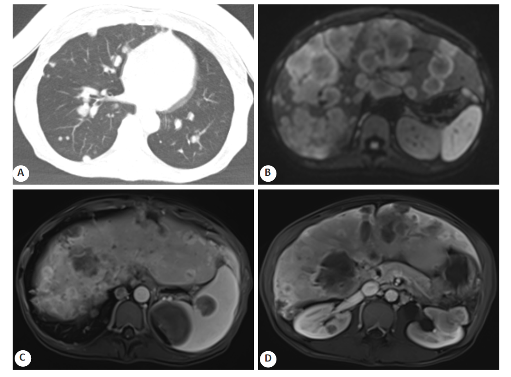
The above images show the baseline imaging assessment of systemic metastases before treatment
A: Chest CT (2026‑08‑31) showing diffuse multiple metastatic nodules in both lungs;
B: Abdominal MRI diffusion‑weighted imaging (2026‑08‑28) showing densely distributed multiple metastatic lesions within the parenchyma;
C: Abdominal MRI T1‑weighted imaging (2026‑08‑28) showing a larger metastatic nodule in the spleen;
D: Abdominal MRI T1‑weighted imaging (2026‑08‑28) showing nodular metastatic lesions in both kidneys.
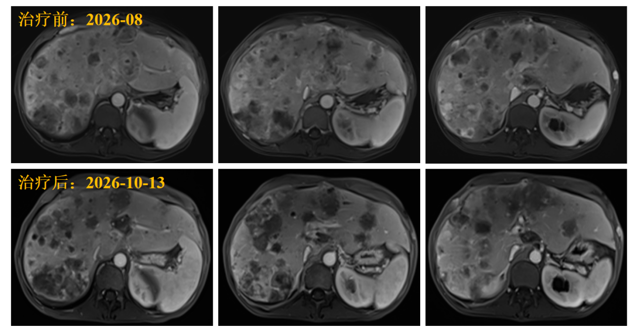
The above images show changes in liver lesions on MRI T1‑weighted imaging before and after treatment of liver metastases.
Compared with the diffusely distributed liver nodules before treatment (August 2026, upper row), follow‑up after treatment (October 13, 2026, lower row) showed changes in contrast/signal characteristics of the liver lesions, with some lesions reduced in size and borders becoming clearer, indicating treatment efficacy.
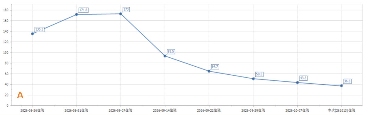
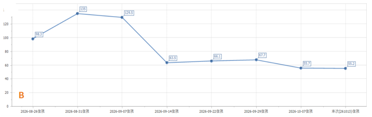
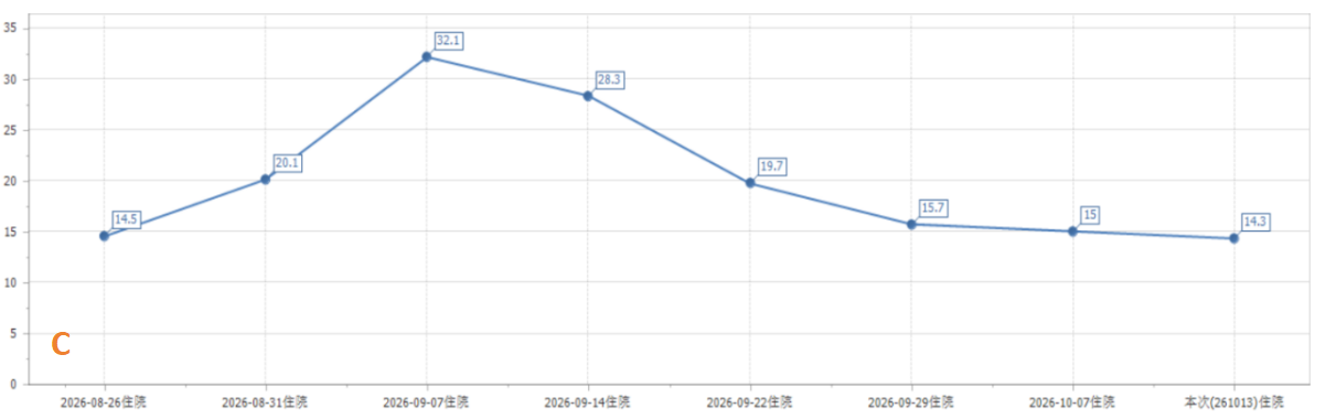
The above images show monitoring changes in hepatobiliary function indicators.
A: Alanine aminotransferase (ALT); B: Aspartate aminotransferase (AST); C: Total bilirubin (TBIL).
Case provided by: Dr. Li Zhenglin
Patient summary: This 37‑year‑old female patient from Singapore developed multiple extensive metastases to the liver, spleen, peritoneum, lymph nodes, and lungs after surgery and postoperative radiotherapy for left pulmonary adenoid cystic carcinoma, classifying as extremely advanced (cT0N0M1c stage IVB). She had previously received multiple lines of pharmacotherapy, all of which were ineffective, with a heavy liver metastatic burden and marked abdominal distension and pain. After heavy (carbon) ion therapy targeting the liver metastatic lesions and biliary system, the patient experienced gradual pain relief, continuous improvement in liver function, and gradual improvement in appetite and mental status during treatment. She tolerated the entire course well with no significant treatment‑related adverse reactions. Post‑treatment follow‑up abdominal MRI showed marked reduction of intrahepatic metastatic lesions compared with before, relief of abdominal pain, and stable general condition. The characteristics of this case are extremely advanced stage, extensive systemic metastases, failure of multiple lines of therapy, and marked symptoms. The advantages of heavy ion therapy lie in its high LET and high RBE, which exert strong radiobiological effects against radioresistant adenoid cystic carcinoma, while the Bragg peak enables precise dose deposition and better protection of surrounding normal liver tissue and the biliary system — thereby demonstrating good efficacy and safety in local control of liver metastases, reduction of tumor burden, relief of pain, and improvement of quality of life. It provides a valuable local treatment option for patients with extremely advanced adenoid cystic carcinoma after failure of multiple lines of therapy.
II. Advantages of Heavy (Carbon) Ions
Physical advantage — "precise blasting" via the Bragg peak: After heavy (carbon) ions enter the body, energy is primarily concentrated and released at the tumor site, forming the Bragg peak. Normal tissues anterior to the tumor receive lower doses, and doses posterior to the tumor drop rapidly — achieving precise strikes that "hit accurately and damage less."
Biological advantage — "armor‑piercing" effect with high RBE: The relative biological effectiveness (RBE) of heavy (carbon) ions is approximately 2 to 5 times that of conventional photons [see references 1‑2], more effectively causing DNA double‑strand breaks in tumor cells; they also have killing effects on hypoxic cells and radioresistant cells.
Spatial advantage — regional control: The treatment range can cover the tumor itself, its invasion pathways, and regional lymph nodes, helping achieve "regional clearance" and reducing "escaped" lesions.
III. Data on Adenoid Cystic Carcinoma Treatment at the Wuwei Heavy (Carbon) Ion Center
As of October 2026, the Wuwei Heavy (Carbon) Ion Center has treated a total of 152 patients with adenoid cystic carcinoma, including 72 males and 80 females, with a mean age of 50.01 ± 15.35 years. Among them, 104 patients had stage IV disease, accounting for 68.42%, most of whom were complex cases with locally advanced disease, postoperative residual disease, or recurrent/metastatic disease.
For stage IV patients, Kaplan‑Meier survival analysis showed a mean survival time of 57.22 ± 6.06 months. The cumulative survival probabilities at 12, 24, 36, 48, and 60 months after treatment were 0.9081, 0.8173, 0.8173, 0.6811, and 0.6811, respectively.
Table 1. Cumulative survival probability of patients with adenoid cystic carcinoma treated with heavy (carbon) ions
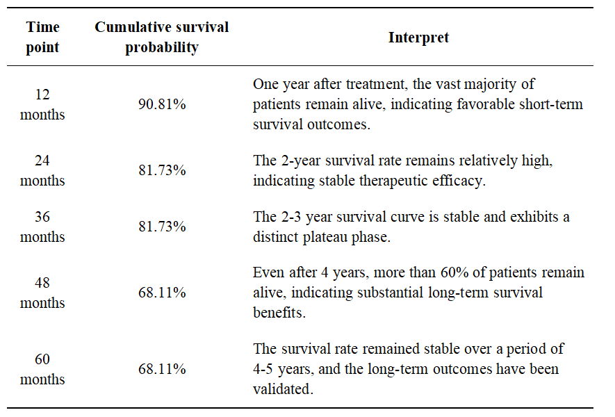
Heavy (carbon) ion therapy for adenoid cystic carcinoma with a high proportion of advanced cases can achieve relatively high short‑ and long‑term survival probabilities, demonstrating good treatment outcomes.
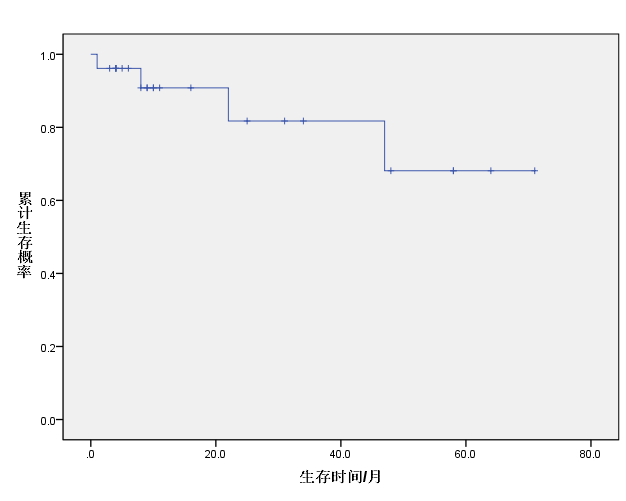
Figure 3. Survival curve of patients with adenoid cystic carcinoma treated with heavy (carbon) ions (0–60 months)
IV. Safety and Tolerability
This case involved palliative and symptom‑relieving treatment. During the entire course, only transient bilirubin fluctuation occurred, which resolved after symptomatic management. No grade 3 or higher acute radiotherapy adverse reactions occurred. The patient's overall tolerability was good, with no treatment‑related adverse events.
V. What to Do After Failure of Multiple Lines of Therapy? Heavy (Carbon) Ion Technology Offers New Approaches to Symptom Relief
Adenoid cystic carcinoma is prone to metastasis to the lungs and other organs. Once metastasis occurs, there are no effective pharmacotherapeutic options. Extremely advanced adenoid cystic carcinoma was once a "tough nut" in cancer treatment. At the Wuwei Heavy (Carbon) Ion Center, domestically produced heavy (carbon) ions — with their high efficacy, low toxicity, and precision — provide a new option for patients with advanced adenoid cystic carcinoma after failure of multiple lines of therapy.
Introduction to the Wuwei Heavy Ion Center
The Wuwei Heavy (Carbon) Ion Center is the clinical application base for China's first heavy (carbon) ion therapy system with complete independent intellectual property rights. It is technically supported by the Institute of Modern Physics, Chinese Academy of Sciences, and clinically operated by Gansu Wuwei Cancer Hospital. Since the first patient was treated in November 2018, as of October 15, 2026, the center has treated more than 3,300 patients, covering more than 100 tumor types, including lung cancer, pancreatic cancer, liver cancer, glioblastoma, nasopharyngeal carcinoma, rectal cancer, and various sarcomas. Heavy (carbon) ions have a strong tumor cell‑killing effect; the relative biological effectiveness of heavy (carbon) ions can reach 2 to 5 times that of conventional radiotherapy and proton therapy, with minimal impact on surrounding normal tissues [1‑2].
As the only medical institution in the world operating two heavy (carbon) ion therapy systems simultaneously, Wuwei Cancer Hospital continues to advance technological innovation and has achieved nine "world‑first" accomplishments, including precision treatment under ventilator control, bladder cancer treatment under precise bladder capacity control, single‑session treatment with surgical implantation of spacers between organs and tumors, and dual Bragg peak heavy (carbon) ion lattice radiotherapy. It has also developed China's first 360° rotatable and height‑adjustable ion radiotherapy chair and completed the world's first batch of multiple cases of cardiac tumor heavy (carbon) ion treatment.
Leveraging 30 research platforms including the CAS Key Laboratory of Heavy (Carbon) Ion Beam Radiation Biology and the Sydney University Biomedical Data Analysis and Visualization Laboratory, the center has conducted more than 30 clinical studies on key tumor types and published nine monographs including the Standard Operating Procedure for Carbon Ion Radiotherapy, gradually building a knowledge system covering physical technology, clinical diagnosis and treatment, and health management. On February 23, 2026, CCTV‑13's Morning News broadcast a special report on the operation of China's first medical heavy (carbon) ion accelerator with independent intellectual property rights; Yang Jiancheng, Deputy Director of the Institute of Modern Physics, Chinese Academy of Sciences, provided an introduction in the program.
[1] Abousaida B, Seneviratne D, Hoppe BS, Ko SJ, Asaithamby A, Cucinotta FA, et al. Carbon ion radiotherapy in the management of hepatocellular carcinoma. J Hepatocell Carcinoma. 2021;8:1169-1182. (Table 1, reproduced with permission from Kavanagh BD. Introduction to clinical radiation oncology. Radiology. 1998;209(1):168.
[2] Apisarnthanarax S, Bowen SR, Combs SE. Proton beam therapy and carbon ion radiotherapy for hepatocellular carcinoma. Semin Radiat Oncol. 2018;28(4):309-320.
Writer: Gong Qinli (Master's degree candidate)
Supervising Physician: Li Zhenglin (Attending Physician)
Data support: Tumor Registry and Follow‑up Office (Chen Xuelian, Li Pengqing)
Reviewed by: Zhang Yanshan (Professor)
Date: October 26, 2026