
A Case Study of Carbon Ion Therapy in Treating Locally Advanced Esophageal Cancer
A Case Study of Carbon Ion Therapy in Treating Locally Advanced Esophageal Cancer
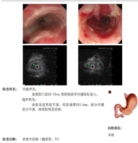
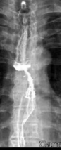
Patient XXX, male, 80 years old, began experiencing difficulty swallowing without obvious cause in February 2022, with progressive worsening. On January 22, 2023, he visited Zhangye People's Hospital, where an upper gastrointestinal contrast study revealed: mid-esophageal stenosis, approximately 5.5 cm in length, with concentric narrowing, mucosal disruption, and irregular filling defects and niches, suggesting mid-esophageal carcinoma (medullary type). On January 25, 2023, gastroscopy at Shanghai XX Hospital showed a circumferential mass protruding into the lumen at 26 cm from the incisors. Pathological examination after biopsy confirmed: (esophageal) squamous cell carcinoma. Upon admission to our hospital, the diagnosis was: 1. Malignant esophageal tumor (mid-esophagus, medullary type, squamous cell carcinoma, cT3N2M0, Stage IVB, KPS score: 90). After multidisciplinary team (MDT) discussion, the treatment plan was determined to be concurrent chemoradiotherapy: 1. Radiotherapy: Photon 40 Gy/20 fractions + carbon ion 16 Gy (RBE)/4 fractions; 2. Chemotherapy: Single-agent oral capecitabine chemotherapy.
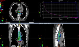
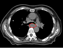
Photon Volumetric Modulated Arc Therapy (VMAT) Plan Diagram
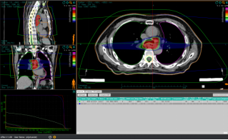
Treatment Planning Image for Carbon Ion Radiotherapy
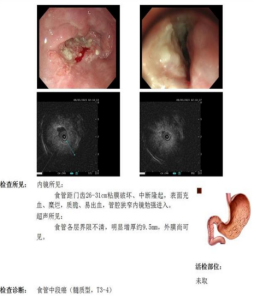
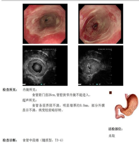
Before Radiotherapy During Radiotherapy After Radiotherapy
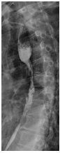
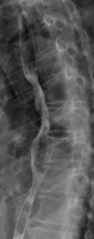
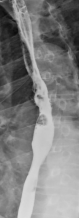
Before Photon Radiotherapy Before Carbon Ion Radiotherapy After Carbon Ion Radiotherapy 1 Month After CarboRadiotherapy
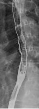
3 Months After Carbon Ion
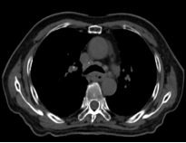
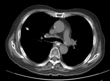
Before Photon Radiotherapy Before Carbon Ion Radiotherapy After Carbon Ion Radiotherapy

