
Case Report on Heavy Ion Therapy for Adenoid Cystic Carcinoma of the Hard Palate
Case Report on Heavy Ion Therapy for Adenoid Cystic Carcinoma of the Hard Palate
Adenoid cystic carcinoma (ACC) is a common malignant tumor in the oral and maxillofacial region and one of the most biologically destructive and unpredictable tumors in the head and neck. It accounts for approximately 28.0% of salivary gland malignancies and is frequently found in the parotid gland and minor salivary glands. Among minor salivary glands, the palatal glands are the most common site of origin. ACC is highly invasive, with a slow clinical progression. The tumor often spreads along nerves (perineural invasion) or through hematogenous dissemination, with the lungs being the most common metastatic site. Lymph node involvement occurs in only about 5-10% of cases.The clinical symptoms of palatal ACC are often atypical, leading to delayed diagnosis until the tumor has grown significantly and extended along the maxillary nerve toward the intracranial region, often rendering surgical resection unfeasible. When radical surgery is not possible, radiation therapy becomes necessary. Literature reports indicate that the 5-year local control and survival rates for surgery or photon radiation therapy range from 25-72% and 25-82%, respectively. In a study of 151 patients with locally advanced ACC involving the oropharynx and paranasal sinuses who underwent carbon ion radiotherapy at NIRS, the 5-year local control rate reached 74%. These results demonstrate that carbon ion radiotherapy provides superior local control compared to intensity-modulated photon radiation therapy alone.Below is a case of locally advanced hard palate ACC. The patient presented with a left hard palate lesion that extended into the naso-cranio-orbital and oral regions, extensively involving surrounding structures. The pons, left optic nerve, and fourth ventricle were compressed, accompanied by widespread cerebral edema. Additionally, there was destruction of the adjacent left mandibular bone, leading to difficulties in mastication, as well as loss of vision, hearing, and smell. Following heavy ion therapy, the patient's symptoms were significantly alleviated.
Medical History: The patient is a 51-year-old female. Chief Complaint: A mass on the hard palate for over 4 years, accompanied by headache and vision loss for 1 year.
Present Illness: The patient first noticed a mass on the hard palate on April 1, 2020, and intermittently received traditional Chinese medicine (TCM) treatment. Subsequently, the mass gradually increased in size. By October 2023, the patient experienced progressive blurring of vision in the left eye, eventually leading to complete vision loss. Despite continued TCM treatment, the symptoms showed no significant improvement. Over the course of the disease, the patient gradually developed localized pain, a sensation of heaviness in the head, left-sided nasal and facial pain, difficulty chewing, and loss of vision, hearing, and smell.On September 11, 2024, the patient visited the Head and Neck Surgery Department of Beijing Cancer Hospital for a biopsy of the palatal mass. The pathological results (Pathology No. 306404) indicated a salivary gland-origin tumor, suspected to be adenoid cystic carcinoma. The patient was prescribed "Anlotinib," but no significant improvement was observed. The patient then came to our hospital for heavy ion therapy.Pre-treatment Cranial MRI Findings: A communicating lesion involving the left hard palate, naso-cranio-orbital, and oral regions was observed, extensively affecting surrounding structures. The pons, left optic nerve, and fourth ventricle were compressed, accompanied by widespread cerebral edema, leading to dilation and fluid accumulation in the ventricular system. Additionally, there was destruction of the adjacent left mandibular bone.
Diagnosis: Adenoid cystic carcinoma of the left hard palate, cT4bN0M0, Stage IVB.
After a thorough evaluation of the condition, a carbon ion treatment plan was formulated. The prescribed doses were as follows:
- CTV (Clinical Target Volume): 52 Gy (RBE) in 13 fractions, 4 Gy (RBE) per fraction.
- GTV (Gross Tumor Volume): 68 Gy (RBE) in 17 fractions, 4 Gy (RBE) per fraction.
The treatment was completed on November 18, 2024. By the 10th treatment session, the patient developed oral mucosal leukoplakia, classified as RTOG Grade 2 acute toxicity. The symptoms were managed with mouth rinses and other supportive treatments, resulting in satisfactory relief.
Before treatment:
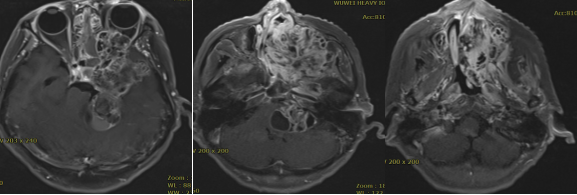
Pre-treatment MRI Findings: A communicating lesion involving the left hard palate, naso-cranio-orbital, and oral regions was observed, extensively affecting surrounding structures. The pons, left optic nerve, and fourth ventricle were compressed, accompanied by widespread cerebral edema, leading to dilation and fluid accumulation in the ventricular system. Additionally, there was destruction of the adjacent left mandibular bone. The findings are highly suggestive of a malignant neoplastic lesion, possibly complicated by localized hemorrhage.
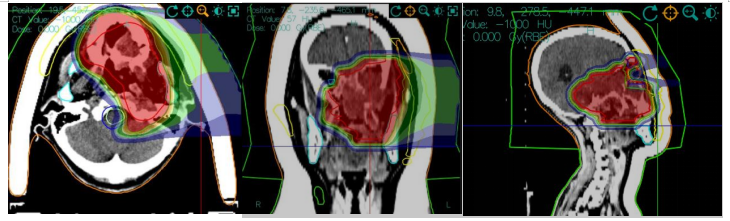
Carbon Ion Radiation Dose Cloud Map
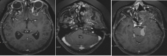
Post-treatment MRI Findings: A communicating lesion involving the left hard palate, naso-cranio-orbital, and oral regions was observed, still extensively affecting surrounding structures. The pons, left optic nerve, and fourth ventricle remain compressed, accompanied by cerebral edema, leading to dilation and fluid accumulation in the ventricular system. Additionally, there is persistent destruction of the adjacent left mandibular bone. However, compared to previous imaging, the tumor size has decreased, and its enhancement has reduced.
Treatment Efficacy Evaluation: The communicating lesion involving the left hard palate, naso-cranio-orbital, and oral regions has shown significant reduction in size compared to previous imaging. The clinical evaluation indicates a Partial Response (PR).
Discussion:
“Carbon Ion Radiotherapy (CRIT)” is an emerging radiotherapy modality in the 21st century. Compared to traditional X-ray radiotherapy, carbon ion therapy offers significant therapeutic advantages and is internationally recognized as one of the most effective cancer treatment methods. It boasts high precision, minimal side effects, reduced damage to normal tissues, high cure rates, and shorter treatment durations. Due to the high energy and strong destructive power of carbon ions, they can precisely target and destroy tumor cells while causing minimal damage to surrounding healthy tissues. This makes carbon ion therapy particularly advantageous for treating certain types of tumors, especially deep-seated tumors and those adjacent to critical organs (such as the optic nerve, brainstem, and lens). Since Japan first applied carbon ion beams to treat malignant tumors in 1994, only a few countries worldwide, including Germany, China, Italy, and Austria, have adopted this technology for clinical use. The Wuwei Heavy Ion Accelerator, China's first independently developed heavy ion tumor treatment device, supports both uniform scanning and spot scanning modes. It was officially introduced into clinical practice in March 2020 and has since been used to treat various cancers, including head and neck malignancies, lung cancer, liver cancer, and pancreatic cancer, accumulating substantial clinical experience.
In the case of hard palate cancer, the patient achieved a “partial response (PR)” following carbon ion radiotherapy, with significant improvement in symptoms such as heaviness in the head and headaches. The treatment was well-tolerated, with mild side effects, demonstrating the efficacy and safety of carbon ion radiotherapy in treating hard palate cancer.

