
Carbon Ion Therapy for Recurrent Glioblastoma
Carbon Ion Therapy for Recurrent Glioblastoma
Glioblastoma (GBM) is the most common primary malignant tumor of the central nervous system in adults, classified as a Grade IV high-grade glioma in the WHO classification system. It is difficult to completely resect surgically and prone to local recurrence postoperatively. Even after standard treatment, recurrence or metastasis may still occur. Approximately 5% of glioblastoma patients experience distant multifocal metastases after standard therapy. This is because the primary tumor is close to the cerebrospinal fluid (CSF) system or the CSF system is opened during surgery, which significantly increases the probability of intracranial tumor dissemination. At the time of recurrence, treatment options are limited, and local therapy remains the most beneficial approach for patients. Carbon ion therapy has certain advantages in this regard. Below is an observation of the therapeutic effect of carbon ion therapy on a patient with local recurrence and intracranial metastasis of glioblastoma after surgery.
Patient Information
Mr. Huang, male, 58 years old.
The patient presented with Broca’s aphasia on August 1, 2022. Brain MRI in August 2022 showed a glioblastoma in the left frontal lobe (45×42×35 mm). On August 25, 2022, the patient underwent surgical treatment. Postoperative pathological examination confirmed the diagnosis of glioblastoma (CNS WHO Grade IV). Immunohistochemistry results are as follows:
• Sample 1: GFAP (+), Olig-2 (+), IDH1 (-), ATRX (+), p53 (scattered +), Ki-67 (approximately 40%), H3K27me3 (+).
• Sample 8: PHH3 (scattered +), Ki-67 (approximately 20%).
Molecular pathology indicated that the MGMT promoter was unmethylated. The patient underwent postoperative radiotherapy (33 fractions), with concurrent temozolomide at 100 mg, followed by sequential temozolomide at 300 mg for 12 cycles.
On October 12, 2023, brain MRI revealed abnormal enhancement in the left frontal-temporal lobe and basal ganglia, as well as abnormal enhancement in the right cerebellar hemisphere, suggesting a high likelihood of metastatic tumors. Physical examination findings included:
• Loss of expressive and communicative abilities.
• Muscle strength of the right upper and lower limbs: Grade 3.
• Muscle strength of the left limbs: Grade 4.
• Muscle atrophy in the right lower limb.
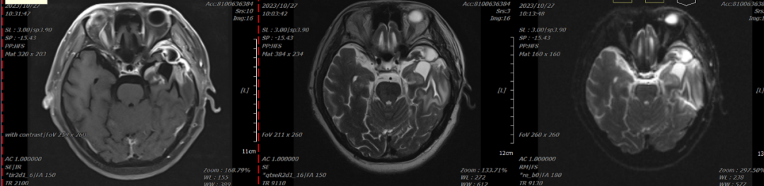
Figure 1: Before treatment, partial absence of the frontal, temporal, and parietal bones, with abnormal signals in the left basal ganglia and left temporal lobe, highly suggestive of postoperative recurrence.
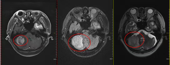
Figure 2: Before treatment, a mass lesion in the right cerebellar hemisphere, with an axial diameter of approximately 3.8 cm × 3.4 cm.
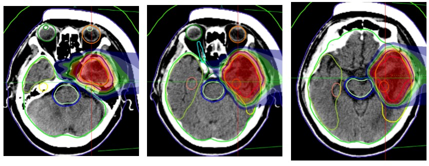
Figure 3: Carbon ion therapy dose cloud map of the left frontal-temporal lobe mass.
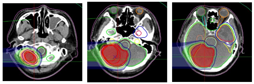
Figure 4: Carbon ion therapy dose cloud map of the right cerebellar lesion.
After treatment, follow-up showed that the cerebellar lesion had shrunk from 3.8 cm × 3.4 cm to 2.1 cm × 1.5 cm. After treatment, the patient's ability to express and communicate, memory, and orientation improved. The muscle strength of the right upper and lower limbs recovered to Grade 3, while the muscle strength of the left limbs returned to normal. No muscle atrophy was observed in the right lower limb.
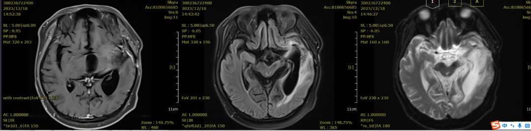
Figure 5: MRI comparison before and after treatment shows abnormal signals in the left basal ganglia and left temporal lobe, with a reduced lesion range and decreased enhancement compared to before treatment.
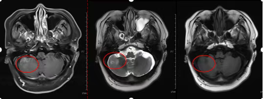
Figure 6: After treatment, a mass lesion in the right cerebellar hemisphere, with an axial diameter of approximately 2.1 cm × 1.7 cm.
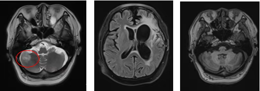
Figure 7: Three months after radiotherapy, a lesion in the right cerebellar hemisphere with an axial diameter of approximately 2.1 cm × 1.5 cm. Enhanced signal in the left temporal lobe decreased.

