
One Case Report of Carbon Ion Radiotherapy for Choroidal Melanoma with Lung Adenocar Cinoma
One Case Report of Carbon Ion Radiotherapy for Choroidal Melanoma with Lung AdenocarCinoma
Choroidal melanoma (CM) is the most common intraocular malignancy in adults, originating from melanocytes in the choroidal stroma. It predominantly affects individuals aged 40–50 and ranks as the second most frequent intraocular tumor. Clinical manifestations vary depending on tumor size and location, with symptoms including vision loss, visual field defects, and vitreous floaters. Extensive retinal detachment can lead to severe vision impairment or blindness. The primary mode of metastasis is hematogenous, with common sites being the liver, skin, gastrointestinal tract, and lungs. Diagnostic methods include fundus fluorescein angiography (FFA), indocyanine green angiography (ICGA), MRI, CT, pathological biopsy, and visual field testing. Choroidal melanoma contains melanin, which exhibits paramagnetic properties, making MRI particularly useful for differential diagnosis. FFA often reveals a "double circulation" phenomenon, characteristic of melanomas that have breached Bruch's membrane. Treatment options include local physical therapies, surgery, and combined therapies. Physical treatments encompass radiotherapy, laser photocoagulation, and photodynamic therapy. Radiotherapy, including brachytherapy and proton therapy, is currently the most commonly used treatment for CM. Although the incidence of CM and lung cancer is relatively independent in the population, the epidemiological characteristics of their coexistence remain unclear. Treatment strategies for CM and lung cancer involve local and systemic approaches, respectively. For CM, treatment options include radiotherapy (e.g., proton beam therapy or stereotactic radiotherapy) and surgery (e.g., enucleation). For lung cancer, treatment depends on tumor stage and molecular characteristics, including surgery, chemotherapy, radiotherapy, targeted therapy, and immunotherapy. When both tumors coexist, treatment strategies must balance the principles of both diseases while considering potential drug interactions and side effects. Carbon ion radiotherapy, with its unique biological effects, has shown efficacy in treating tumors resistant to photon therapy. This report presents a case of carbon ion radiotherapy for choroidal melanoma coexisting with lung adenocarcinoma, focusing on its effectiveness and adverse reactions.
Clinical Data
Ⅰ. Patient Information
A 41-year-old female presented with "blurred vision in the right eye for 20 days, worsening with a sense of visual obstruction for 10 days." The patient reported sudden onset of blurred vision in the right eye 20 days prior, accompanied by a noticeable visual obstruction in the upper temporal field, affecting both near and distant vision.
Ⅱ. Examinations
• Orbital MRI (Right Eye):A mound-shaped abnormal signal with mild enhancement was observed on the posterior upper and lower walls of the right eyeball, suggestive of melanoma.
• Chest CT: A mass in the right lower lobe was identified, likely primary lung cancer.
•Lung Biopsy: Histopathology revealed invasive acinar adenocarcinoma. Immunohistochemistry results: C-erbB-2(-), CK7(+), Ki-67(index=25%), NapsinA(+), P63(-), TTF-1(+).
•Diagnosis:
1. Right choroidal melanoma.
2. Right lung invasive acinar adenocarcinoma, cT3N0M0 Stage IIb (AJCC 8th edition), KPS: 90.
3. Partial retinal detachment in the right eye.
Ⅲ. Diagnosis and Differential Diagnosis
Diagnosis
1. Right choroidal melanoma.
2. Right lung invasive acinar adenocarcinoma, cT3N0M0 Stage IIb (AJCC 8th edition), KPS: 90.
3. Partial retinal detachment in the right eye.
Differential Diagnosis
1. Inflammation: Choroidal inflammation with associated retinal detachment or granulomas may mimic melanoma due to pigment epithelial hyperplasia. However, inflammatory lesions often exhibit surrounding edema and hemorrhage, which are less common in melanoma.
2. Choroidal Nevus:Benign melanocytic lesions are typically smaller, less elevated, and rarely cause visual field defects. Fluorescein angiography shows no fluorescence.
3. Macular Disciform Degeneration: Hemorrhagic or serous lesions in the macula may resemble melanoma, but subretinal hemorrhage and drusen are distinguishing features. Ultrasound and FFA can aid in differentiation.
4. Choroidal or Subretinal Hemorrhage: Hemorrhages secondary to hypertension, diabetes, or vascular diseases can mimic melanoma. Examination of the contralateral eye and ultrasound are helpful in diagnosis.
Ⅳ.Treatment
1. Carbon Ion Radiotherapy Planning:
• Head Positioning:Supine position with head-neck-shoulder immobilization. CT scan with 2 mm slice thickness covering the entire brain.
• Chest Positioning: Prone position with vacuum cushion and thermoplastic immobilization. CT scan with 3 mm slice thickness from the thoracic inlet to the inferior liver margin, including plain, contrast-enhanced, and 4D sequences.
• Target Delineation: MRI images were fused with CT scans for precise delineation of the choroidal and lung lesions.
2.Treatment Plan:
• Right Choroidal Tumor: GTV: Right choroidal tumor; PGTV: GTV with 2 mm margin. Dose: 70 Gy(RBE)/5 fractions.
• Lung Tumor: GTV: Right lung lesion; CTV: GTV with 8 mm margin; PTV: GTV and CTV with 5 mm margin. Dose: CTV: 45 Gy(RBE)/15 fractions; GTV boost: 27 Gy(RBE)/3 fractions; Total dose: 72 Gy(RBE)/15 fractions.
3. Efficacy Evaluation:
• Vision and Intraocular Pressure: Monitored before, during, and after treatment (Table 1). Post-treatment MRI showed slight tumor enlargement with no light perception. One-month follow-up MRI revealed stable disease (SD) (Figure 4).
• Lung Lesion: Pre-treatment size: 4.7 cm × 5.2 cm; Post-treatment size: 4.2 cm × 3.5 cm. Evaluation: SD (Figure 5).
4. Acute Adverse Effects:
• Mild right eye edema was observed after three treatment sessions, with no other acute complications.
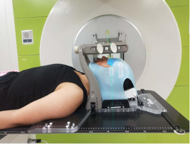
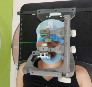
Figure 1: Positioning Fixation (Custom Head Frame Fixator)
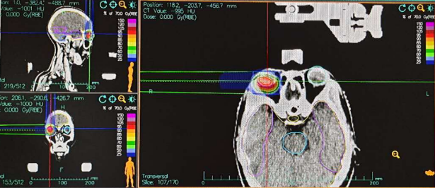
Figure 2: Radiotherapy Target Volume for Right Choroidal Tumor GTV: Right choroidal tumor; PGTV: GTV with a 2 mm margin. Dose: 70 Gy(RBE)/5 fractions
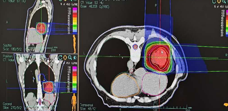
Figure 3: Radiotherapy for Right Lung Lesion CTV: 45 Gy(RBE)/15 fractions; Tumor Boost: 27 Gy(RBE)/3 fractions; Total Dose: 72 Gy(RBE)/15 fractions
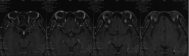
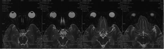
Figure 4: Abnormal mound-shaped signal on the posterior upper and lower walls of the right eyeball, showing mild enhancement. Melanoma cannot be ruled out. A nodule is observed on the posterolateral wall of the right eyeball, exhibiting short T1 and T2 signals, with high signal on DWI
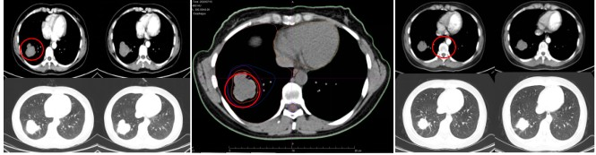
Figure 5: Pre-treatment size of the lung lesion: 4.7 cm × 5.2 cm; Post-treatment size on chest CT after 1 month: 4.2 cm × 3.5 cm
Table 1: Pre- and Post-Treatment Eye Examination Results
| Oculus Dexter | Oculus Sinister | The Examination Results of Dexter | ||
|---|---|---|---|---|
| VOD | IOP | VOS | IOP | |
| HM/30cm | 9.8 mmHg | 1.0 | 16.4 mmHg | A mass approximately 3 PD in size is observed in the superotemporal quadrant of the right eye. The retina below appears bluish-gray and elevated. The optic disc is faintly visible, and the macular structure is absent. |
| Light Perception (Right) | 13.9 mmHg | 1.0 | 18.8 mmHg | The retina below the right eye appears bluish-gray and elevated, with the optic disc structure absent and the macular structure disappeared.。 |
| HM/BE | 11.0 mmHg | 0.8 | 18.2 mmHg | The right eye exhibits cortical opacity of the lens, with clear boundaries of the optic disc. Retinal elevation is observed inferiorly, and the superior tumor has reduced in size compared to previous findings. The retina shows degeneration and atrophy, with pigment deposition. The macular structure is indistinct, and the light reflex is absent. |
V. Follow-up and Outcomes
Evaluation of Visual Acuity and Intraocular Pressure: Visual acuity and intraocular pressure were assessed before, during, and after treatment. Post-treatment MRI revealed a slight increase in tumor size, with no light perception. One month after treatment, MRI showed no significant change in tumor size compared to pre-treatment, and the efficacy was evaluated as stable disease (SD).
Evaluation of Pulmonary Lesions: Before radiotherapy, the size of the pulmonary lesion was 4.7 cm × 5.2 cm. One month after treatment, chest CT showed a reduction in size to 4.2 cm × 3.5 cm, also evaluated as SD.
Acute Adverse Effects: Mild edema of the right eyeball was observed after three treatment sessions, with no other acute injuries.
Discussion
Malignant melanoma is a type of malignant tumor derived from melanocytes, commonly found in the skin but also occurring in mucous membranes, the choroid of the eye, and other sites. It is characterized by early metastasis, high mortality, and resistance to conventional radiotherapy. Choroidal melanoma and lung cancer are both highly aggressive malignancies originating from the choroid and lung tissue, respectively. Although their primary sites differ, an increasing number of patients are found to have both conditions in clinical practice. Treatment for choroidal melanoma includes local therapies such as radiotherapy and surgery, as well as systemic therapies like targeted therapy and immunotherapy. Lung cancer treatment involves surgery, radiotherapy, chemotherapy, and targeted and immunotherapies. For patients with both conditions, treatment strategies must be individualized, considering the staging of both tumors, the patient’s overall condition, and potential therapeutic interactions. In this case, the patient underwent concurrent heavy ion radiotherapy for both choroidal melanoma and lung cancer. Although the incidence of melanoma in China is lower than in Western countries, it is rising significantly. Heavy ion therapy provides a more effective treatment option for patients with malignant melanoma. In 2001, the National Institute of Radiological Sciences (NIRS) in Japan initiated clinical trials of heavy ion radiotherapy for ocular melanoma, which concluded in 2004. As expected, heavy ion radiotherapy proved to be safe and effective. By February 2013, 127 patients with locally advanced or unfavorably located ocular melanoma had undergone heavy ion therapy, with 5-year survival and eye preservation rates of 80.8% and 93.1%, respectively. The local tumor control rate and intraocular recurrence-free rate were 96.4% and 92.3%, respectively. Compared to conventional photon therapy, carbon ions exhibit superior physical and biological properties. While photon beams exhibit exponential dose attenuation in tissue, carbon ion beams release relatively low energy along their path, with a significant energy release at the end, forming a Bragg peak. Beyond the Bragg peak, the dose is minimal. In radiobiology, carbon ions have a higher linear energy transfer (LET), directly causing 70% of DNA double-strand breaks in tumor cells, making them effective against cells in all phases of the cell cycle. In summary, for melanomas that are difficult to completely resect or respond poorly to conventional radiotherapy, heavy ion therapy offers better local control and overall survival rates, with no significant radiation-related adverse effects. As a local treatment modality, heavy ions will play a crucial role in managing malignant melanoma. Therefore, the clinical application of heavy ions for treating refractory tumors like malignant melanoma urgently needs to be promoted. It is believed that in the near future, heavy ion radiotherapy for melanoma will have broader clinical applications.

