
Heavy ion therapy for Bulbar glioma
Introduction
Medullary glioma, also known as bulbar glioma, refers to a glioma occurring in the medulla oblongata. It belongs to the category of brainstem gliomas, which typically originate in the pons, followed by the midbrain, medulla oblongata, cerebral peduncles, and cervical spinal cord. Brainstem gliomas are further classified into diffuse, exophytic, cervicomedullary, and focal intrinsic types. The medulla oblongata, located at the lowest part of the brainstem, primarily controls respiratory functions and also governs other vital activities. Symptoms of medullary tumors can be specific or non-specific. Patients with medullary tumors include both adults and children. When a brainstem tumor develops within the medulla oblongata, patients commonly experience symptoms such as unsteady gait, hoarseness, difficulty swallowing, tongue deviation to one side, and facial numbness. In severe cases, even the patient's breathing during sleep can be affected, manifesting as abnormal breathing rhythms.
The medulla oblongata is a region densely packed with nerves, containing fiber tracts that connect the spinal cord and the brain, as well as cranial nerves such as the glossopharyngeal, vagus, accessory, and hypoglossal nerves. It houses vital centers responsible for basic life functions like heartbeat, respiration, and digestion. Due to its complex anatomical relationships and deep-seated location, surgical intervention in this area is highly challenging. Radiotherapy, as a non-invasive treatment modality, can precisely target the tumor site, effectively eradicating cancer cells. The advanced nature of heavy ion radiotherapy lies in its dual physical and biological effects: the dose of heavy ions is primarily deposited at the end of their range (Bragg Peak), making it the radiation therapy with the least damage to healthy tissues and minimal side effects. With a high relative biological effectiveness (RBE), heavy ions induce DNA double-strand breaks and have a low oxygen enhancement ratio, making them suitable for treating hypoxic tumors. The small divergence of the ion beam allows for high-precision treatment, making it an ideal option for difficult-to-treat tumors that are inoperable, resistant to conventional radiation, or recurrent after other treatments.
Here is a case of a patient with a malignant tumor in the medulla oblongata. Due to the lesion's location in the medulla oblongata, specifically a space-occupying lesion in the right part of the brainstem, the adjacent fourth ventricle was compressed, leading to symptoms such as headache, dizziness, coughing while drinking, nystagmus, right-sided facial paralysis, numbness on the right side of the face, and weakness in the left lower limb. After undergoing heavy ion therapy, the patient's symptoms significantly improved.
Medical History:
- Patient: Male, 32 years old
- Chief Complaint: Weakness in the left lower limb for over 5 months, diagnosed with a malignant brainstem tumor for over 1 month.
Present Illness:
The patient experienced weakness in the left lower limb in August 2024 without any obvious cause. After receiving traditional Chinese acupuncture treatment, the symptoms showed no significant improvement. Two months ago, the patient developed headaches and dizziness. One month ago, there was a noticeable decline in hearing on the left side. On November 26, 2024, a cranial MRI revealed a "space-occupying lesion in the brainstem." Subsequently, on December 19, 2024, the patient sought treatment at Beijing Tiantan Hospital, where a "stereotactic brain lesion resection" was performed. Postoperative pathology indicated a diffuse glioma with ATRX loss.
Immunohistochemistry Results:
- Ki-67: Approximately 10% positive
- IDH-1: Negative
- ATRX: Loss
- H3K27M: Negative
- H3K27me3: Positive
- CD34: Positive
- BRAF: Positive
- CD20: Negative
- CD3: Negative
Supplementary Immunohistochemistry Results:
- NF: Positive
- Syn: Positive
- MAP-2: Positive
- P53: Overexpression
- CD68: Negative
- MSH2: Positive
- MSH6: Positive
- MLH1: Positive
- PMS2: Positive
Postoperative Condition:
After surgery, the patient experienced dizziness, right-sided facial paralysis, and numbness on the right side of the face. Heavy ion therapy was subsequently administered.
Diagnosis:
- Medullary Astrocytoma, IDH-mutant, MGMT-methylated
- WHO Grade II
- KPS Score: 70
This case highlights the complexity of treating brainstem gliomas, particularly in critical areas like the medulla oblongata. The use of heavy ion therapy post-surgery demonstrates a multidisciplinary approach to managing such challenging tumors, aiming to improve patient outcomes and quality of life.
Treatment Plan: Photon + Carbon Ion Therapy
Target Delineation:
- GTV (Gross Tumor Volume): The visible medullary lesion on MR T2 flair imaging.
- CTV (Clinical Target Volume): GTV with a 1.5 cm margin.
First Course: Photon Radiotherapy
- Prescription Dose: 44 Gy in 40 fractions.
- Dose per Fraction: 1.1 Gy.
- Frequency: Twice daily.
Second Course: Heavy Ion (Carbon Ion) Radiotherapy
- Prescription Dose: 15.5 Gy (RBE) in 5 fractions.
- Target: GTV.
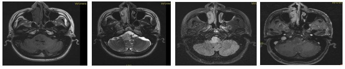
Pre-treatment MRI: A space-occupying lesion in the right part of the brainstem, measuring 2.2 × 2.0 cm, with compression of the adjacent fourth ventricle, highly suggestive of glioma.
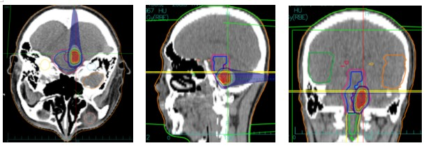
Carbon Ion Therapy Dose Distribution Map
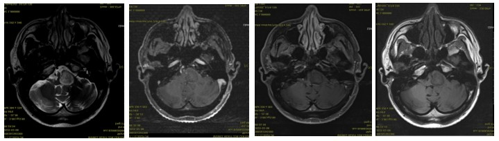
Post-treatment MRI: A space-occupying lesion with enhancement in the right part of the brainstem, accompanied by compression of the adjacent fourth ventricle, showing little change compared to the previous scan (2025-01-10). Efficacy evaluation: SD (Stable Disease).
Discussion:
Carbon ion radiotherapy (CIRT) is an emerging radiotherapy modality in the 21st century. Compared to traditional X-ray radiotherapy, carbon ion therapy offers significant advantages in treatment efficacy and is internationally recognized as one of the leading cancer treatment methods. It is characterized by high precision, minimal side effects, reduced damage to normal tissues, high cure rates, and shorter treatment durations. Due to the high energy and strong destructive power of carbon ions, they can precisely target and destroy tumor cells while causing minimal damage to surrounding healthy tissues. This makes carbon ion therapy particularly advantageous for treating specific types of tumors, especially deep-seated tumors and those located near critical organs (such as the optic nerve, brainstem, and lens).
Since Japan first applied carbon ion beams to treat malignant tumors in 1994, only a few countries, including Germany, China, Italy, and Austria, have adopted this technology for clinical use. The Wuwei Heavy Ion Accelerator, China's first independently developed heavy ion tumor treatment device, supports both uniform scanning and spot scanning modes. It was officially put into clinical use in March 2020 and has since been used to treat various cancers, including head and neck malignancies, lung cancer, liver cancer, and pancreatic cancer, accumulating substantial clinical experience.
In this case, the short-term efficacy evaluation of carbon ion radiotherapy for a patient with a malignant medullary tumor was assessed as SD (Stable Disease). The patient's symptoms, such as headache, dizziness, coughing while drinking, nystagmus, right-sided facial paralysis, numbness on the right side of the face, and weakness in the left lower limb, showed improvement compared to their condition at admission. The treatment was well-tolerated, with no significant adverse reactions observed during the course of therapy. This case confirms the efficacy and safety of carbon ion radiotherapy in the treatment of malignant medullary tumors.

