
Female patient, 69 years old, locally advanced esophageal cancer
[Typical Case] Carbon Ion Therapy for a Patient with Locally Advanced Esophageal Cancer
Clinical History:
A 69-year-old female developed dysphagia on October 10, 2023, progressing to tolerating semi-liquid diets. On December 26, 2024, PET-CT at our hospital revealed:
- Focal wall thickening and mass formation in the lower thoracic esophagus
- Increased metabolic activity
- Corresponding luminal stenosis Findings consistent with esophageal cancer.
Endoscopic Ultrasound (EUS):
- At 31-35 cm from incisors: Mucosal irregularity with semi-circular elevated lesion Ulceration with white slough Luminal stenosis Blurring of mucosal/submucosal/muscularis propria layers Layer thickening: 11.8–14.8 mm
- Pathology: Poorly differentiated squamous cell carcinoma.
Diagnosis: Squamous cell carcinoma of lower esophagus, Metastatic mediastinal lymph nodes, Stage cT3N1M0 (IIIB)
Treatment:
Following multidisciplinary team (MDT) discussion:
- Concurrent carbon ion radiotherapy + capecitabine chemotherapy
- Post-7 carbon ion sessions (EUS re-examination):Lesion thickness: 12.9–14.2 mm
- Treatment plan redesigned → 8 additional carbon ion sessions
- Post-treatment EUS:Lesion thickness reduced to 5.0–6.0 mm
- Outcome: Dysphagia resolved; tolerating soft diet.
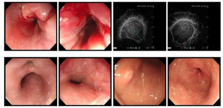
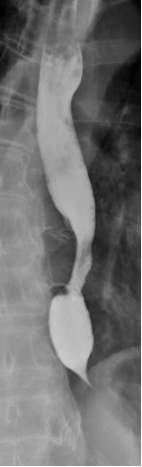
Figure 1: Pre-treatment EUS (2024-12-28) *At 31-35 cm from incisors:*
Mucosal irregularity, semi-circular elevated lesion with ulceration and white slough. Luminal stenosis. Blurred mucosal/submucosal/muscularis propria layers. Thickness: 11.8–14.8 mm.
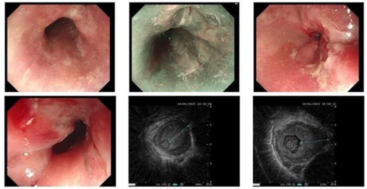
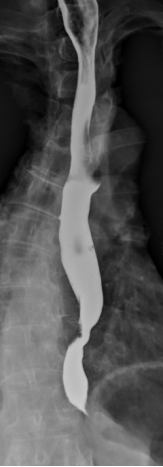
Figure 2: Interim EUS (2025-01-14) *At 31-35 cm from incisors:*
Mucosal irregularity, semi-circular elevated lesion, luminal stenosis. Blurred layers. Thickness: 12.9–14.2 mm.
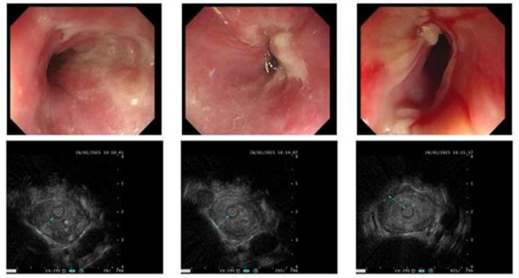
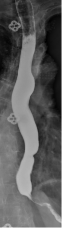
Figure 3: Post-carbon ion therapy EUS (2025-01-26)*At 27-31 cm:* Mucosal irregularity with localized pallor.
*At 31-35 cm:* Mucosal irregularity, semi-circular elevated lesion, luminal stenosis. Blurred layers. Thickness: 5.0–6.0 mm.
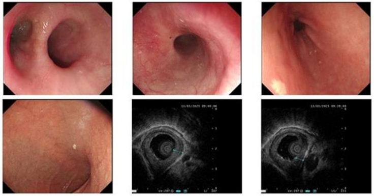
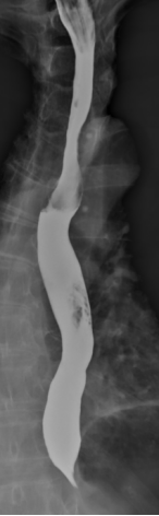
Figure 4: 2.5-month post-therapy EUS (2025-03-13)
*At 29-31 cm:* Mucosal irregularity with pallor. Blurred layers. Thickness: 3.7–4.4 mm.
2025.01.05 2025.01.15 2025.02.05 2025.06.25

