Hydrogel Spacer-Assisted Heavy Ion Radiotherapy for Sacrococcygeal Chordoma
Hydrogel Spacer-Assisted Heavy Ion Radiotherapy for Sacrococcygeal Chordoma
Chordoma originates from embryonic notochord remnants, with more than half of all cases occurring in the sacrococcygeal region. These tumors are locally aggressive and have an insidious early presentation. As they grow, they can compress the cauda equina, leading to constipation and difficulty with urination. Chordomas are relatively radioresistant to photon therapy and often require high‑dose radiotherapy (proton/carbon ion doses typically reaching 60–74 Gy(RBE)) for tumor control. Given the proximity of sacrococcygeal lesions to the anterior wall of the rectum, in some cases the tumor may share the rectal wall or even encase the bowel. The rectal tolerance dose is significantly lower than the curative tumor dose; exceeding this limit can lead to radiation proctitis, bleeding, and perforation. However, reducing the radiation dose around the intestine may increase the risk of tumor recurrence. To enable curative‑dose radiotherapy for sacrococcygeal chordoma while protecting the rectum, we present the CT‑guided technique of hydrogel spacer implantation in the recto‑tumor space to assist heavy ion radiotherapy. Before radiotherapy, an absorbable tissue spacer is implanted between the lesion and the rectum to physically create a safe distance. The hydrogel spacer, composed primarily of polyethylene glycol (PEG) hydrogel, is an absorbable biomaterial. It is injected as a liquid precursor and, within 10 seconds of reaching the target space, absorbs water and solidifies in situ. It maintains stable morphology for approximately 3 months, is partially absorbed by 6 months, and is completely absorbed and naturally excreted through urine by 9 months, with no long‑term retention. The key advantage is that it physically separates the anterior rectal wall by approximately 1–2 cm, significantly reducing the high‑dose volume to the rectum (e.g., V70), thereby lowering the risk of radiation proctitis, bleeding, and urgency.
Our hospital, in collaboration with the Department of Radiation Oncology at Peking University Third Hospital, performed the first CT‑guided hydrogel spacer implantation in the recto‑tumor space to assist heavy ion radiotherapy for sacrococcygeal chordoma in China.
Pelvic MRI and CT of the sacrococcygeal chordoma revealed bone destruction of the S4‑Co1 vertebrae with an associated soft tissue mass. The lesion was closely adherent to the posterior wall of the rectum, with significant rectal distension due to gas accumulation, further thinning the bowel wall and nearly obliterating the natural space between the tumor and the rectum. After Professor Wang Junjie's ward round and multidisciplinary consultation, the CT‑guided rectal spacer hydrogel implantation procedure was performed by Professor Guo Fuxin, with the assistance of Chief Physician Li Xiaojun and Deputy Chief Physician Lu Jing. [Case provided by Dr. Shen Yamei]
Pre‑implantation Imaging
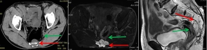
Figure 1. Before hydrogel spacer implantation, the lesion is closely adherent to the posterior wall of the rectum, with significant rectal distension due to gas accumulation (Red arrow: sacrococcygeal lesion; Green arrow: rectum)
Hydrogel spacer is percutaneously implanted into the perirectal space under CT guidance, which physically separates the lesion from the rectum. This technique ensures adequate radiation dose delivery to the target lesion while reducing the radiation dose absorbed by the rectum.
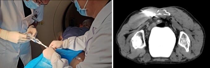
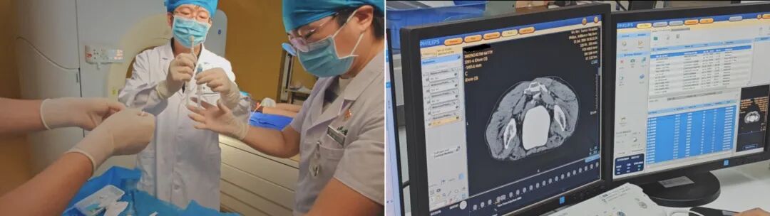
Figure 2. CT‑guided implantation of hydrogel spacer in the rectal space
Radiographic Comparison Before and After Hydrogel Spacer Implantation
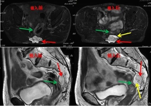
Figure 3. Radiographic comparison before and after hydrogel spacer implantation: The hydrogel spacer displaces the anterior rectal wall forward by approximately 8.9 mm, establishing a stable physical barrier (Red arrow: sacrococcygeal lesion; Green arrow: rectum; Yellow arrow: hydrogel spacer)
Comparison of Rectal Radiation Dose Before and After Spacer Implantation
Following hydrogel spacer implantation, we re‑planned the carbon ion radiotherapy plan. Key pre‑ and post‑implantation rectal dose metrics for this case are summarized below:
- Rectal Dmax: 67 Gy(RBE) → 25 Gy(RBE), a reduction of 42 Gy(RBE), bringing it from a high‑risk range to within safe limits.
- Rectal V70: Approximately 8% → approximately 0%, eliminating the high‑dose volume and significantly reducing the risk of radiation proctitis.
- Rectal V60: 10 mL → <1 mL, meeting clinical tolerance criteria (V60 < 1 mL).
- Tumor prescription dose: Post‑spacer, a safe dose of 70.4 Gy(RBE) carbon ion radiotherapy was delivered, meeting the curative dose requirement for chordoma.
What were the previous methods for implanting spacer materials?
Early methods of spacer placement included:
Open surgical placement (earliest method):
An incision was made in the abdomen or perineum to expose the presacral space under direct vision, allowing manual placement of the spacer. Major disadvantages included significant trauma, prolonged hospitalization, high cost, and elevated risks of infection and bleeding.
Laparoscopic placement:
Absorbable sheets were placed and sutured in position via laparoscopy. While less invasive than open surgery, this approach still required general anesthesia, surgical team involvement, and high costs. It also remained technically challenging in cases of large tumors.
Surgical placement of hemostatic gauze pads:
During surgery, absorbable hemostatic gauze was packed into the space between the tumor and the bowel. However, this method had drawbacks including uneven thickness, unpredictable absorption rates, lack of standardized protocols, and the need to remove the gauze on the same day after placement, resulting in surgical trauma.
Transperineal hydrogel injection:
Hydrogel was injected via ultrasound‑guided puncture through the perineum into the anterior rectal space. However, ultrasound has limited visualization of deep presacral structures, the puncture pathway is long, and the gel tends to distribute unevenly, making it difficult to achieve consistent and complete coverage.
CT‑Guided Hydrogel Spacer Implantation – Key Advantages
Precision: Visible needle tip and gel
CT‑based real‑time tomography clearly delineates the sacrum, tumor margins, rectal wall, blood vessels, and nerves. The needle tip position is accurate to the millimeter level, and the entire puncture trajectory is fully visible. Gel distribution is monitored in real time during injection to ensure uniform coverage of the tumor‑bowel interface with no gaps. Immediate post‑procedure CT confirms gel position, spacer thickness, and excludes rectal wall infiltration or needle‑track hematoma, allowing any issues to be addressed on the spot.
Minimally invasive: A small puncture replaces a large incision
CT‑guided percutaneous placement eliminates the need for incisions or general anesthesia, and patient recovery is significantly faster than with any surgical spacer placement method. This avoids complications associated with large incisions, including infection, bleeding, and intestinal adhesions. It is particularly advantageous for elderly patients and those with multiple comorbidities.
Controllable: Dose‑verification closed‑loop
Radiotherapy plans are compared before and after implantation. DVH curves are used to confirm that rectal V60, V70, and Dmax all meet tolerance criteria before radiotherapy is initiated. The gel is not simply placed and assumed to be effective; placement is verified, and only after verification does radiotherapy proceed. Referencing Shiba et al.'s dose‑volume study: after spacer placement, rectal Dmax was reduced from 67 to 25 Gy(RBE) — a reduction of approximately 42 Gy(RBE) — enabling patients who were previously ineligible due to rectal dose constraints to safely receive curative doses.
Safe: Robust evidence base
Absorbable hydrogel spacers (such as SpaceOAR) are supported by strong safety evidence and are certified by FDA, CE, and NMPA, and recommended by NICE. A phase III RCT in prostate cancer (222 patients) demonstrated a 74% reduction in rectal V70, with a 0% vs. 5.7% incidence of grade ≥2 rectal toxicity at 3 years, and natural hydrolytic absorption within 3–6 months with no residual material. In a carbon ion radiotherapy case for sacrococcygeal chordoma, preoperative absorbable spacer placement reduced rectal Dmax from 67 to 45 Gy(RBE) (a reduction of 42 Gy), enabling curative‑dose treatment in a previously inoperable case and further validating its safety and efficacy in complex anatomical sites.
Technical Takeaways from This Case
CT guidance is the "gold standard" for deep‑site puncture: In this case, significant rectal distension and an irregular tumor‑bowel interface made ultrasound‑guided placement nearly impossible. CT's cross‑sectional imaging capability penetrates gas interference, allowing the operator to clearly visualize needle‑tip displacement and gel distribution down to the millimeter.
Spacer gel transforms "inoperable" into "treatable": A 42 Gy(RBE) reduction in Dmax is not just a numerical change — it represents a qualitative shift from "inevitable damage" to "safe and controllable" treatment. This case demonstrates that even in the most challenging anatomical configurations, curative doses can be safely delivered when the appropriate technique is applied.
Minimally invasive value is significant: Compared with open surgery or laparoscopy, CT‑guided percutaneous placement requires only local anesthesia, minimal trauma, and fewer adverse effects. This is particularly important for middle‑aged and elderly patients with underlying comorbidities.
Dosimetric verification is indispensable: Implantation alone does not guarantee success. In this case, immediate post‑procedure CT was performed to verify spacer thickness, followed by re‑planning and DVH comparison to confirm that all rectal dose parameters met tolerance criteria before radiotherapy was initiated. This closed‑loop workflow — "implantation → verification → planning → radiotherapy" — is the core safeguard for patient safety.
[Writer: Shen Yamei; Reviewer: Li Xiaojun; Department: First Department of Radiotherapy (Lanzhou Campus)]
Writer: Shen Yamei, Li Xiaojun
First Review: Liu Qiong
Second Review: Guo Yishan
Third Review: Cai Qinghua