Encyclopedia of Images | Claustrophobia Meets MRI, How to Break the Deadlock?
Encyclopedia of Images | Claustrophobia Meets MRI, How to Break the Deadlock?
Imagine being in a narrow, confined, and roaring space—fear washes over you like a tidal wave, and breathing grows labored. This is precisely the dire predicament that claustrophobic patients face during traditional MRI scans.

However, at the Lanzhou Heavy Ion Center, a technological innovation is quietly transforming their fate: the 3.0T large-bore high-end positioning MRI.
Recently, a patient long plagued by claustrophobia successfully underwent an examination here. This achievement represents not only a triumph of personal courage but also a breakthrough jointly forged by technological innovation and humanistic care.
Claustrophobia: The Invisible Hurdle on the Path to Medical Imaging
Claustrophobia is a common anxiety disorder characterized by intense fear, a sense of loss of control, and even physical symptoms such as palpitations, sweating, and shortness of breath when individuals are exposed to enclosed or crowded spaces. Traditional MRI scanners feature narrow bores (typically around 60 cm in diameter), lengthy scan durations (which may extend to 15–45 minutes or more), and are accompanied by continuous, sometimes loud noise. These compounded factors create a nearly insurmountable barrier for patients with claustrophobia. Consequently, many such individuals refuse or postpone necessary imaging examinations, potentially delaying the diagnosis and treatment of critical illnesses.
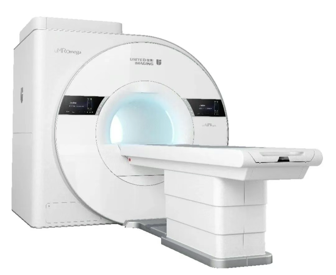
As the only 3.0T large-bore high-end positioning MRI scanner in urban Lanzhou, it was specifically developed to shatter this "invisible barrier":
Technical Advantage
01 Large-aperture Design
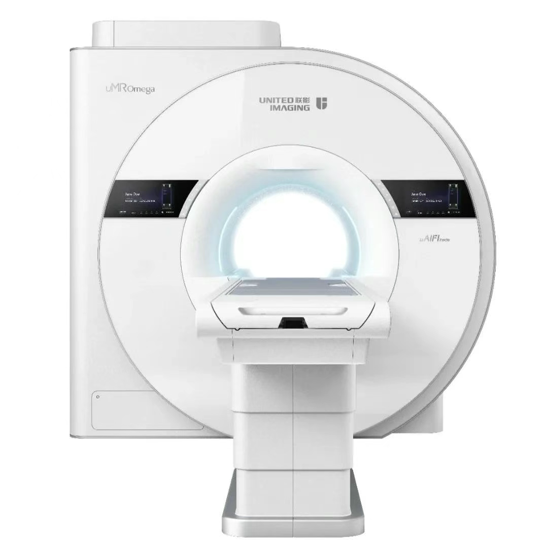
Adopting a 97cm ultra-large inner diameter net magnet assembly, its 75cm bore is 25% larger than that of conventional MRI scanners, providing a more comfortable examination experience for special groups with specific imaging needs such as pregnant women and obese individuals.
02 Ultra-high-performance Gradient System
Equipped with the industry's first single-unit gradient power amplifier boasting a peak power of 3.5 megawatts, this system effortlessly handles advanced clinical and research applications, even high-precision radiotherapy simulation and localization, thereby highlighting the distinct advantages of heavy ion therapy MRI localization.
03 Intelligent Imaging Technology
Equipped with United Imaging's second-generation acceleration platform, OptiStar UCS2.0, and ACS Intelligent OptiStar Imaging Technology, this system integrates compressed sensing technology, parallel acquisition technology, and other advanced techniques to enable whole-body imaging within 100 seconds, significantly reducing examination time.
04 Advanced Coil Technology
Distinctive Magic Carpet Coil, quilt-like in its gentle and comfortable feel, boasts advantages of high signal-to-noise ratio (SNR) and high resolution, providing patients with enhanced scanning comfort.
05 Life Perception Technology
Pioneering millimeter-wave radar life perception technology to monitor patients' respiratory conditions, supporting high-precision monitoring of small-amplitude respiration, enabling real-time display of patients' vital signs, and ensuring high-quality MRI images and patient safety.
Clinical Application
01 Tumor Screening and Diagnosis
Whole-body diffusion-weighted imaging (DWI) can serve as a radiation-free whole-body "PET-CT-like" imaging modality, enhancing the visualization of lesions—particularly improving the detection rate of malignant tumors and their metastases. Meanwhile, it features ultra-high temporal resolution within 3 seconds, enabling more precise quantitative analysis of DCE data for tumor characterization, prediction, and evaluation of treatment efficacy.
02 Neurological Examination
Adaptive optimized variable flip angle matrix3D sequence combined with LightSpeed Imaging 2.0 can display all 12 pairs of cranial nerves in a single scan, significantly improving the display rate and anatomical details of fine cranial nerves, and assessing the degree of nerve invasion. Additionally, it enables functional imaging modalities such as perfusion, nerve fiber bundle (DTI) imaging, resting-state imaging, and task-based blood oxygen level-dependent imaging (BOLD).
03 Cardiovascular System Examination
It can clearly display cardiac structure and function, which is of great value for the diagnosis of myocardial lesions, valvular heart disease, etc. The intelligent vascular plaque analysis post-processing engine can automatically perform vascular wall analysis and intelligent plaque analysis, providing a basis for the prevention and treatment of cardiovascular diseases.
04 Abdominal Examination
Sub-second abdominal 4D multi-phase dynamic enhancement (T-UCS) with LightSpeed dynamic enhancement imaging supports up to 32x acceleration, achieving 0.5s/phase sub-second temporal resolution. This enables complete acquisition of the entire dynamic enhancement process including early, mid, and late arterial phases, allowing precise delineation of the extent and nature of abdominal lesions.
05 Musculoskeletal System Examination
The application of the UCS2.0 platform enables free zooming of local images, while 3D MPR reconstruction enhances joint imaging to be clearer and more three-dimensional, significantly improving the diagnostic rate and diagnostic accuracy of bone and joint lesions. The new MAPS cartilage quantification platform, combined with UCS3D imaging, can precisely visualize minute cartilage lesions.
Humanistic Care: Accompanying Through the Entire Process, Melting the Ice in Hearts
We deeply understand that technology is the foundation, and care is the bridge. For special patients, we have developed a personalized service plan called "Reassuring Examination":
Before the Examination: Thorough communication is key. Medical staff will provide detailed explanations of the equipment features (with emphasis on the advantages of the large bore), the examination process, and possible sounds that may be heard. Patients are invited to enter the examination room in advance to familiarize themselves with the environment, and even undergo a brief adaptive experience.
During the Examination: Family members are allowed to accompany in the safe zone, providing strong psychological support; technicians maintain communication through the intercom system throughout the process, informing the progress and soothing emotions at any time; patients can wear dedicated noise-canceling headphones/earplugs; more comfortable positioning pads are used; and the examination can be paused at any time based on the patient’s feelings.
Patient’s Voice: From "Impossible" to "I Did It"
"In the past, just the thought of having an MRI would make me feel suffocated and my hands and feet go cold. But the doctors and nurses here are especially understanding. They first showed me that 'big machine,' and the space was really much larger than I imagined. They even let me lie in it to try it out. During the examination, someone kept talking to me, telling me what to do at each step. Knowing I was scared, they also allowed my family member to stand next to me so I could see him... It felt completely different from before. I finally completed an examination I never dared to imagine!" shared Ms. Wang, a patient who successfully underwent the examination, with excitement. This examination provided crucial imaging evidence for her subsequent possible treatment.
A Friendly Reminder:
If you or your family members/friends feel anxious about conventional imaging examinations due to claustrophobia, body size, or other reasons, please proactively inform the medical staff. The Medical Imaging Department of our hospital is willing to work with you to explore solutions, utilizing advanced technologies such as large-bore MRI and personalized services to help you overcome obstacles and complete necessary health examinations with peace of mind.
Lanzhou Heavy Ion Center
Gansu Wuwei Cancer Hospital is a state-owned public Grade III, Class A specialized tumor hospital. The Lanzhou Heavy Ion Center, operated by the hospital, is the second China-developed heavy ion clinical treatment center in the country. It houses the second domestically-developed China-made heavy ion therapy system, which commenced clinical operations on November 15, 2024. To date, the center has completed over 230 cases of photon and heavy ion therapy, covering cancers such as lung cancer, pancreatic cancer, liver cancer, and glioblastoma.
The Lanzhou Heavy Ion Center has established an elite talent ecosystem, anchored by a 50-member expert workstation led by globally renowned authorities. This includes Shen Wenjiang, Professor Emeritus of Radiation Oncology at Peking University Health Science Center, and Xu Bo, Standing Committee Member of the Chinese Society for Radiation Oncology. Dr. Wang Junjie, Chairman of the Chinese Society for Radiation Oncology, serves as the hospital’s President, providing robust technical leadership for heavy ion therapy.
Equipped with cutting-edge medical facilities, the center features:
- Varian VitalBeam medical linear accelerator
- Elekta Infinity four-dimensional image-guided linear accelerator
- 3.0T MRI for simulation positioning
- GE 256-slice high-end CT
- Varian-Iridium 192 afterloader
These advanced systems ensure high-precision and efficient treatment workflows.
Guided by the vision of delivering comprehensive, lifelong cancer care, the hospital offers multidisciplinary services through departments such as:
- Radiation Oncology
- Medical Oncology/Hematology
- Surgical Oncology
- Integrated Traditional Chinese and Western Medicine Rehabilitation
- Critical Care Medicine
- Gastroscopy Center
- Imaging, Laboratory, and Blood Banking
The center integrates photon therapy, chemotherapy, heavy ion therapy, surgery, and advanced diagnostics to provide holistic treatment solutions.