
What can PET-CT detect?
As the name implies, PET-CT combines two imaging technologies—PET (Positron Emission Tomography) and CT (X-ray Computed Tomography). In clinical practice, these complementary imaging modalities enable physicians to examine the human body's internal structures from different perspectives.
You may be more familiar with CT. Simply put, it involves taking X-ray images from multiple angles and then synthesizing them to create a three-dimensional image. For example, the world’s first X-ray image shows the flat skeletal structure of Röntgen’s wife’s hand. Today, with CT technology, we can reconstruct every cross-sectional layer of a person’s brain—from the base to the top.
But have you noticed? What CT provides is structural information (what organs look like), not functional details (how organs are working). Take these brain CT images, for instance—we can see the physical structure, but we can’t tell which areas are "active."
That’s where PET comes in. Like CT, PET also involves an image synthesis process. However, unlike CT, which captures images through X-rays, PET requires the subject (usually a patient in clinical settings) to ingest a tracer substance tagged with radioactive markers, and then tracks the distribution of these radioactive markers within the body.
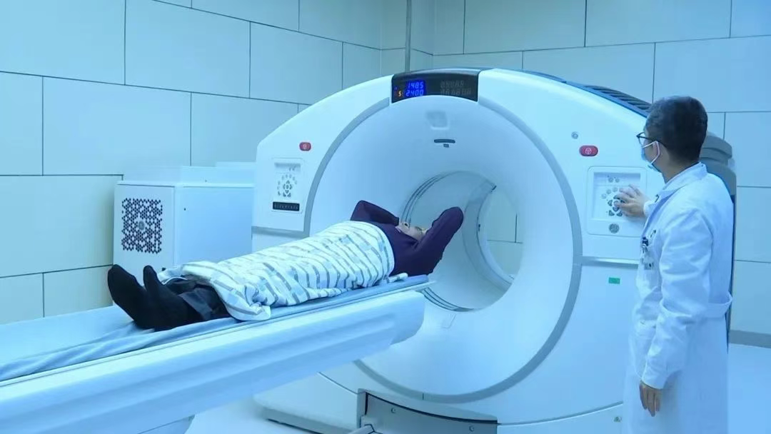
In common PET imaging, patients are administered a radioactive tracer called "fluorodeoxyglucose (FDG)"—its name already hints at its similarity to glucose. Indeed, this tracer not only shares a similar molecular structure with glucose but also follows a comparable distribution pattern within the human body after administration.
It is precisely this characteristic that allows FDG to function as a diagnostic tool: when introduced into the body, tissues or cells with higher glucose uptake will accumulate more of this tracer. Since the tracer emits radiation, its distribution can then be captured by imaging equipment (PET). The result? We can visualize which organs or tissues in the patient’s body have "consumed" more glucose.
You might wonder: Why do we need to know which tissues in the body "consume" more sugar?
Here’s the key: Glucose is a critical energy source for the human body. When PET reveals glucose distribution patterns, it simultaneously reflects the metabolic activity levels of organs, tissues, and even cells at the microscopic level (think of it this way: the more "work" a cell does, the more glucose it "eats").
In oncology, this principle becomes particularly powerful. Cancer cells typically exhibit abnormally high metabolic rates—they divide and proliferate uncontrollably, demanding excessive energy ("eating" more to fuel their growth). Furthermore, many tumors develop a distinct metabolic preference for glucose. These characteristics make PET imaging an invaluable tool for detecting and visualizing tumors.

When PET and CT are combined for cancer imaging, CT provides precise information about the tumor's location, size, and structural characteristics, while PET reveals metabolic activity levels. The fusion of these two modalities offers unique diagnostic insights that no single technology can achieve alone. This is why PET-CT plays a pivotal role in cancer diagnosis and treatment.
For instance, during diagnostic evaluations, physicians leverage the contrasts between malignant and healthy tissues visible on PET-CT scans to facilitate accurate diagnoses. In treatment monitoring, comparing pre- and post-treatment PET-CT images enables clinicians to assess tumor response and determine therapeutic efficacy.
Consequently, PET/CT demonstrates unequivocal advantages in detecting primary tumors, metastases, and recurrent lesions at early stages—earning its reputation as the "life radar" for tracking tumor progression.
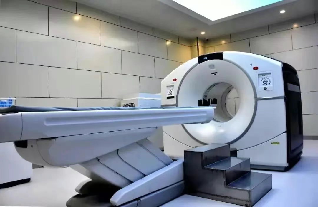
The PET-CT Center at Wuwei Medical Science Academy Tumor Hospital in Gansu Province has introduced the GE Discovery 710 Super Iterative PET-CT—the most advanced 256-ring LBS crystal PET/128-slice spiral CT system in China. This state-of-the-art equipment is the fourth PET-CT unit in Gansu Province and the only one installed in a prefecture-level hospital across the province. As a flagship super-iterative device launched by GE Healthcare, it sets the global benchmark for diagnostic imaging, delivering superior sensitivity and specificity compared to conventional PET/CT systems.
Key technical highlights include:
- Unparalleled spatial resolution of 1.8mm, enabling significantly improved detection of tiny lesions
- Dramatically reduced whole-body scan time, while simultaneously lowering radiation exposure for patients
- Optimal diagnostic outcomes that have earned it the reputation as the "crown jewel of modern medical technology"
The Center is equipped with a first-class radiopharmaceutical laboratory and a medical cyclotron system capable of producing various disease-specific tracers, including ¹¹C, ¹³N, and ¹⁸F. The synthesized radiotracers achieve purity levels exceeding national standards and reaching world-class quality, ensuring precise diagnostic results for patients. As a result, our PET-CT Center has become the only facility in Gansu Province capable of performing both ¹¹C and ¹⁸F imaging—solidifying its leadership in advanced oncologic diagnostics.

