
【Popular Science Mini-Lesson】What Exactly Are Calcifications in an Ultrasound Report?
【Popular Science Mini-Lesson】What Exactly Are Calcifications in an Ultrasound Report?
During an ultrasound examination, the report may mention "calcifications." Most people do not have a clear understanding of this term, which can cause significant psychological stress. Today, we will briefly introduce several common types of calcifications.

What are calcifications?
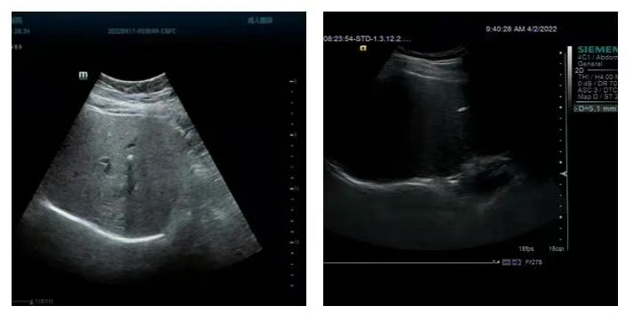
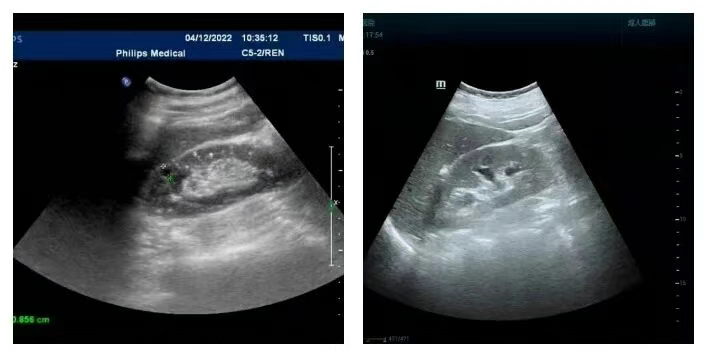
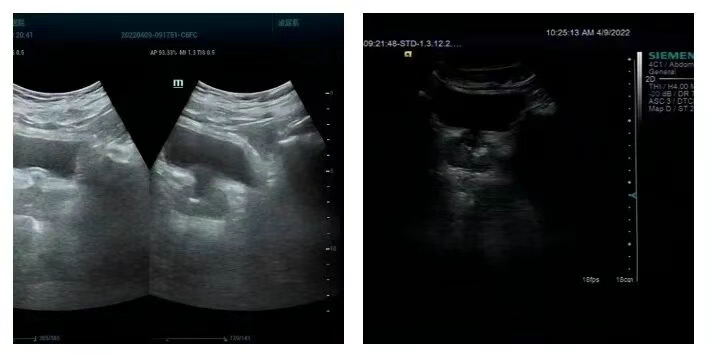
Calcifications refer to calcium deposits detected in an organ via color Doppler ultrasound or CT imaging, appearing as stone-like strong echoes or high-density shadows. Common types include liver calcifications, prostate calcifications, kidney calcifications, and lung calcifications

Formation of Calcifications
Calcifications are generally caused by inflammation and tuberculosis. For example, after pulmonary tuberculosis is cured, it can form lung calcifications, similar to scars. Calcified spots in the liver might be due to calcification in the bile duct walls, often resulting from inflammation, tuberculosis, or possibly past trauma, hemorrhage, abscesses, or granulomas forming "scar-like" calcifications.
Typically, these indicate the lesion is stable or healed and does not require special treatment.
Several Common Types of Calcifications
1.Liver CalcificationsThese appear as stone-like strong echoes or high-density images in the liver on color Doppler ultrasound or CT scans. They are more common in people aged 20-50, with equal occurrence in men and women. Usually, it's a single calcification, more frequent in the right lobe than the left, and rarely appear in both lobes simultaneously. Many conditions can lead to liver calcifications, including:
• Intrahepatic bile duct stones, which is the most common factor;
• Chronic inflammation or trauma within the liver;
• Parasitic infections;
• Benign or malignant liver tumors and calcified intrahepatic metastases;
• Congenital development, where calcifications can form in the fetus in utero, often associated with congenital malformations, with an incidence of 0.057%
Kidney Calcifications
This refers to the hardening and calcification of local kidney tissue, usually acquired gradually over time due to recurrent local inflammation causing calcium deposition or various other reasons. Detected calcifications are essentially calcium deposits within the kidney and generally cannot disappear on their own, with only a small amount being absorbed by the tissue. If they do not progress or cause pathological changes, they usually do not lead to other impacts. Kidney calcifications should be checked regularly. In most cases, they are simply special variations resulting from necrotic cells after normal metabolism. As the body undergoes daily metabolism, the necrosis of some cells is normal. After necrosis, if local circulation is poor, calcium salts can deposit, forming calcified plaques that appear as bright spots similar to stones on ultrasound
Prostate Calcification and Fibrosis
This is the scar left after prostatic inflammation heals and is a precursor to prostatic stones. Prostate stones are often accompanied by chronic prostatitis, and these changes can usually be seen through ultrasound examination. Due to the particular structure of the prostate, there are generally no particularly effective treatments for calcification and stones once they occur. Bacteria can breed on prostate calcifications/fibrosis or stones, which can be a cause of recurrent prostatitis and should not be ignored. Calcified spots in the prostate are mostly caused by chronic inflammation or are prostatic stones. They usually do not cause symptoms and do not require treatment.
Through the above introduction, we believe you now have a basic understanding of calcifications. In the future, if you encounter such descriptions in your examination reports, there is no need to panic. However, since the human body is constantly changing, it is still important to remind everyone to maintain close observation and get timely re-examinations.
