Foam Test: Unveiling the "Hidden Alarm" for Heart Health
"Frequent headaches, dizziness, or even unexplained strokes? The hidden 'culprit' might be lurking in your heart!"
Today, we’re talking about a test that may seem "magical" but is incredibly important—the foam test. Dubbed the "detective of heart health," it can uncover invisible risks that the naked eye can’t see.
What exactly is the foam test? Who needs it? Today, we’ll break it all down for you in one clear article!
01 Foam Test: Tracing Bubbles to Uncover Hidden Heart Risks
The full name of the foam test is Contrast Transcranial Doppler (C-TCD). It involves injecting saline solution containing tiny microbubbles into the patient’s antecubital vein (elbow vein), then instructing the patient to perform a Valsalva maneuver (hold their breath and strain) or cough. Using a TCD microemboli detection system, doctors observe how these microbubbles travel through the veins.
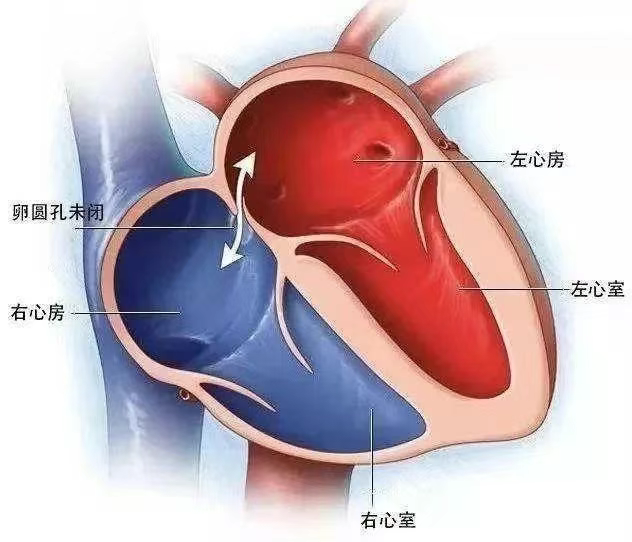
Under normal circumstances, the bubbles are absorbed in the pulmonary circulation and cannot be detected by TCD as microemboli. However, if a patent foramen ovale (PFO, an opening between the heart’s atria that didn’t close properly after birth) exists, the bubbles can pass from the right atrium through the unsealed foramen ovale into the left atrium, and subsequently flow into the brain’s blood vessels. Under ultrasound monitoring, these bubble signals can then be captured and analyzed.
Principle:
Physiological saline is oscillated to produce tiny microbubbles → injected into a vein → observed via ultrasound whether the bubbles "abnormally migrate" (i.e., bypass the lungs and enter systemic circulation).
Key Objective:
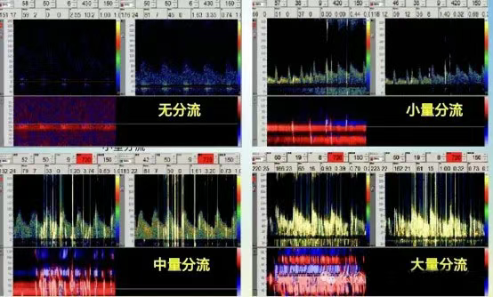
To screen for right-to-left cardiac shunts (such as patent foramen ovale). Such shunts may allow venous thrombi, gases, or other substances to directly enter the brain, potentially causing strokes or migraines.
The transcranial Doppler (TCD) test is widely used for clinical screening of right-to-left shunts due to its high sensitivity, safety (non-invasive), and ease of repetition. This helps improve diagnostic accuracy for identifying the underlying cause, and provides important evidence for developing individualized prevention and treatment plans.
Follow-up Recommendation:
If the result is positive, further evaluation with transesophageal echocardiography (TEE) or cardiac CT is needed to determine the shunt volume and exact location. The physician will then assess whether interventional closure treatment is required.
Address:
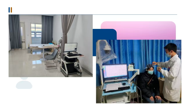
Room 428, Department of Cardio-Cerebral Function Examination, 4th Floor, Outpatient Building, Central Campus, Wuwei Cancer Hospital
Address:
Room E303, Department of Cardio-Cerebral Function Examination, 3rd Floor, Outpatient Building, Heavy Ion Campus, Wuwei Cancer Hospital