
PET-CT: The Key Tool for Clinically Precise Staging of Oncological Diseases
PET-CT: The Key Tool for Clinically Precise Staging of Oncological Diseases
PET-CT is currently one of the world's most advanced nuclear medicine molecular imaging devices, known in Chinese as Positron Emission Computed Tomography/X-ray Computed Tomography.

PET-CT, also known as hybrid imaging or molecular imaging, is a tracer imaging technology.It tracks the activity of specific biological substances in the human body by using positron-labeled tracers. The system employs multi-layer, ring-shaped detectors to detect photons emitted by the tracers outside the body. The acquired optical data are then processed by a computer to generate anatomical images along with corresponding physiological parameters, revealing the condition of target organs or diseased tissues for diagnostic purposes.
In addition to combining the individual functions of PET and CT, PET-CT uniquely integrates their images—fusing metabolic data from PET with anatomical details from CT. This enables the simultaneous assessment of lesion metabolism and structure, facilitating early disease detection while significantly improving diagnostic accuracy. As a result, PET-CT has been hailed as a powerful tool for precise clinical staging of malignant tumors.
Precision Clinical Staging
1. According to currently internationally recommended diagnostic standards, approximately 20% of surgical patients are confirmed to have benign nodules through postoperative pathology;
2. In cases where the goal is curative treatment, focal and distant metastases are often detected shortly after surgery.
Therefore, precise clinical staging of malignant tumors holds significant clinical importance for formulating rational treatment plans and evaluating prognosis.
Case 01
Patient with Cardia Carcinoma
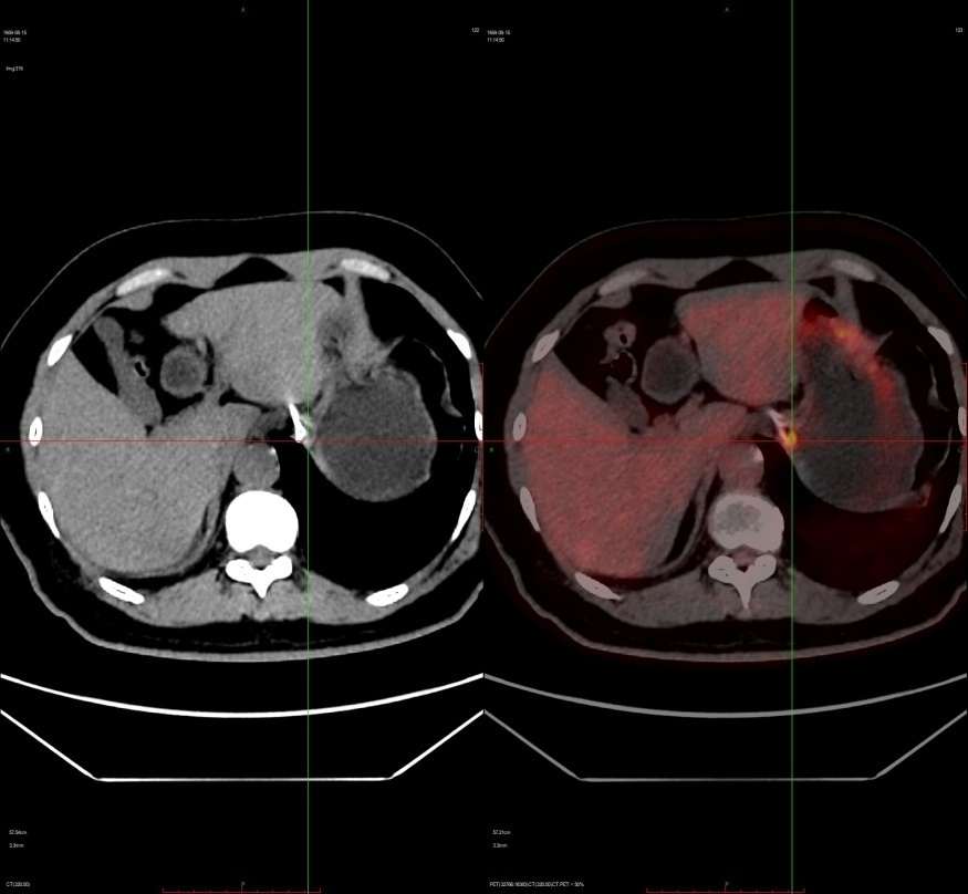
Figure 1 Local lesion in the cardia
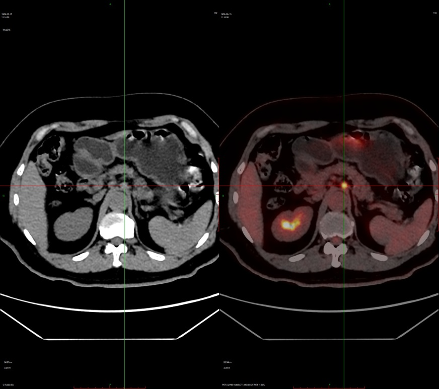
Figure 2 Metastatic lymph nodes anterior to the abdominal aorta
Precision Clinical Staging: T1bN1M0, Stage IB
Case 02
Patient with Pancreatic Carcinoma
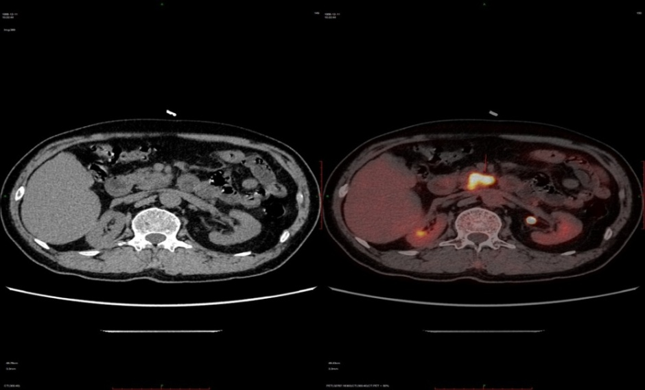
Figure 1 Focal lesion in the pancreas
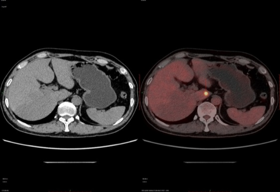
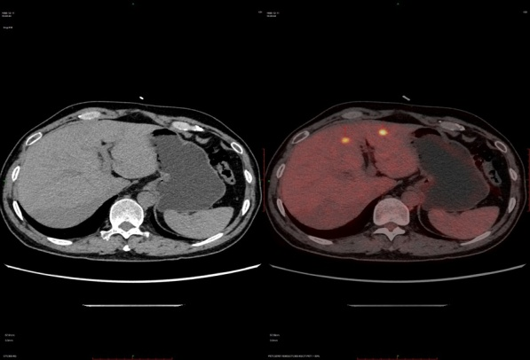
Figure 2 Multiple metastatic lesions in the hepatic region
Precision Clinical Staging:
Pancreatic carcinoma (2cm < maximal diameter < 4cm) with multiple hepatic metastases, T2N0M1, Stage IV
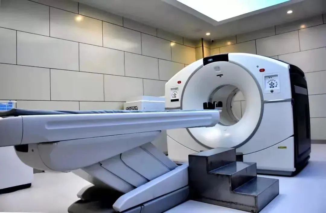
The PET-CT Center at Wuwei Academy of Medical and Science Cancer Hospital in Gansu Province has introduced the GE Discovery 710 Clarity Super Iterative PET-CT from the United States, which is currently the most advanced 256-ring LBS crystal PET/128-slice spiral CT in China. This device is the fourth PET-CT in Gansu Province and the only one installed in a prefecture-level hospital across the province. As a super iterative device launched by GE Healthcare that represents the cutting edge of global technology, it offers significantly higher sensitivity and specificity compared to conventional PET/CT systems. Featuring an unprecedented 1.8mm spatial resolution, the system greatly enhances detection capabilities for minute lesions while substantially reducing whole-body scan time and radiation exposure for patients, delivering optimal diagnostic results. It has been hailed as the pinnacle of modern medical imaging technology. The Center boasts state-of-the-art radiopharmaceutical facilities, equipped with a medical cyclotron system capable of producing various disease-specific 11C, 13N, and 18F tracers required for different diagnostic applications. The purity of the produced radiopharmaceuticals exceeds national standards, reaching world-class levels, thereby ensuring highly accurate diagnoses for patients. Our PET-CT Center is the only facility in Gansu Province capable of performing both 11C and 18F imaging examinations.
Contact us
Consultation phone: Director Li Ke (+86 13689357660)
Medical address: PET-CT Center, Heavy Ion Hospital District, Wuwei Academy of Medical and Science

