
“Zapping Leg Clots: The Vascular Scavenger’s Strike”
Interventional Treatment of Lower Extremity Venous Thrombosis: The "Vascular Scavenger" Protecting Lives
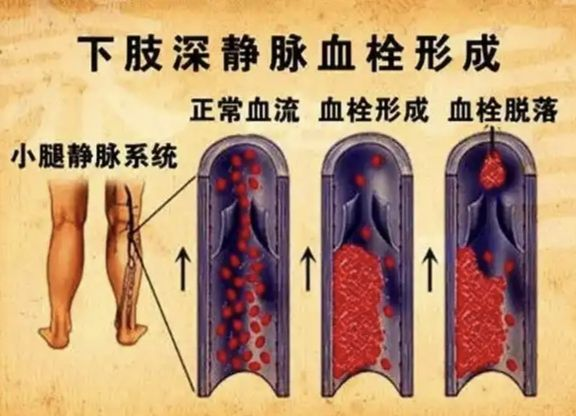
Lower extremity venous thrombosis (DVT) is a disease caused by abnormal blood clotting within the deep veins of the lower extremities. It can lead to swelling and pain in the affected limb, and in severe cases, even cause a life-threatening pulmonary embolism (PE) due to the dislodged thrombus.
I. Core Methods of Interventional Treatment
1.Inferior Vena Cava Filter Implantation: The "Umbrella" Intercepting Life-Threatening Clots
This is the crucial first step of interventional treatment. Through puncture of the femoral vein or jugular vein, a filter shaped like the scaffold of an umbrella is implanted into the inferior vena cava (the largest vein in the human body). This filter is used to intercept the thrombi that detach from the lower extremities, preventing them from migration into the pulmonary artery and causing a pulmonary embolism.
Applicable Scenarios: Extensive deep vein thrombosis (involving both the thigh and the calf), patients with anticoagulation contraindications or those for whom anticoagulation treatment is ineffective, and patients at high risk of pulmonary embolism (such as those with malignancies or prolonged immobilization patients).
High Efficiency: Mechanical thrombectomy can quickly remove the thrombus. Combined with thrombolytic drugs, it can significantly shorten the course of the disease (in some patients, the swelling subsides 2 days after the operation).
Safety: The success rate of the filter in intercepting thrombi exceeds 95%, greatly reducing the mortality rate of pulmonary embolism.
Minimally Invasive: The operation only requires local anesthesia, with a small wound (such as a pinhole). It can be completed within 30 minutes, and patients can be mobilized early after the operation.
2.Thrombectomy: Directly "Declotting" the Vasculature
Mechanical Thrombectomy/Thrombus Fragmentation: The thrombus is fragmented and then aspirated through a catheter (such as using a high-speed rotating catheter to crush the thrombus), which is suitable for acute and relatively large thrombi.
Catheter-Directed Thrombolysis: The thrombolytic catheter is directly inserted into the interior of the thrombus, and drugs such as urokinase are locally injected to dissolve the thrombus. Compared with systemic thrombolysis, this method requires less drug dosage, has lower side effects, and is more effective.
3.Combined Treatment: Employing Multiple Approaches to Improve the Therapeutic Effect
In clinical practice, filter implantation is often combined with thrombectomy. For example, a filter is first placed to prevent pulmonary embolism, and then the thrombus is removed through thrombolysis or thrombectomy, fundamentally solving the problem of blood flow obstruction.
II. Typical Case
The patient, a 73-year-old female, developed swelling of the left lower limb 3 days before admission without any obvious precipitating factors. She also experienced soreness, fatigue, and coldness of the limb, but she did not pay attention to it and did not receive any diagnosis or treatment. Subsequently, the swelling of the left lower limb progressively worsened, and her movement was significantly limited. The color Doppler ultrasound of the lower extremity veins in an external hospital showed thrombosis in the left external iliac vein, common femoral vein, deep femoral vein (branch segment), upper and middle segments of the superficial femoral vein, and intermuscular veins. She was admitted to the Department of Cardiac Intervention on February 12, 2025, with the diagnoses of "1. Lower extremity venous thrombosis; 2. Iliac vein compression syndrome". After admission, "inferior vena cava filter implantation + lower extremity venous catheter thrombolysis" was performed. After the operation, "heparin sodium injection" was given for anticoagulation, and "urokinase for injection (400,000 units) was pumped intravenously twice a day" for thrombolysis. After thrombolytic treatment, the reexamination of the lower extremity venography showed that the thrombus burden was significantly reduced. Then, iliac vein stent implantation was performed. After the operation, rivaroxaban was continued for anticoagulation treatment.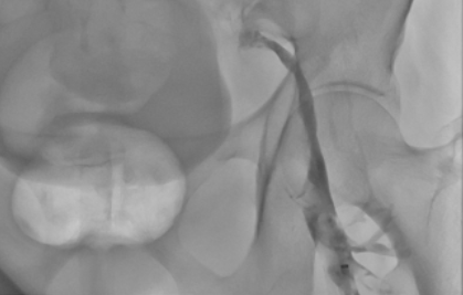
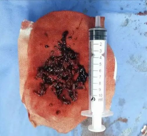
Lower Extremity Venography Thrombus Aspirated during the Operation
The re-examination of the color Doppler ultrasound 1 month after the operation showed: Thrombosis in one intermuscular venous plexus of the left lower limb, and within normal limits were found in the B-mode imaging and color Doppler flow signals of the left external iliac vein, the confluence segment of the great saphenous vein, the common femoral vein, the superficial and deep femoral veins, the popliteal vein, and the posterior tibial vein of the left lower limb.
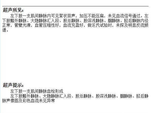 Results of the Venous Color Doppler Ultrasound after the Operation
III. Clinical Recommendations from Experts
The Best Treatment Time: Intervention within 48 to 72 hours after the onset of the disease has the optimal outcomes and can significantly reduce sequelae.
High-Risk Groups: prolonged immobilization patients, cancer patients, pregnant women, and peripartum women should be vigilant against symptoms such as swelling of a single leg and skin warmth, and seek medical attention in a timely manner.
Avoiding Misunderstandings: Do not use unorthodox treatments such as therapeutic phlebotomy, as it may aggravate vascular damage or induce thrombosis.
Interventional treatment provides an efficient and safe solution for patients with lower extremity venous thrombosis through minimally invasive means. Early identification of symptoms and timely medical treatment are crucial.
The minimally invasive surgeries mainly carried out by the Department of Cardiology and Intervention are as follows:
(1) Heart and Great Vessels
Transcatheter Closure of Congenital Heart Diseases (Atrial Septal Defect, Ventricular Septal Defect, and Patent Ductus Arteriosus); Percutaneous Coronary Intervention (PCI) for Coronary Heart Disease, Pacemaker Implantation for Bradyarrhythmia, and Radiofrequency Ablation for Tachyarrhythmia.
Endovascular Exclusion of Aortic Dissection/Aneurysm with a Stent Graft; Endovascular Exclusion of Abdominal Aortic Aneurysm with a Stent Graft; Stent Implantation for Aortic Ulcer and Intramural Aortic Hematoma.
(2)Minimally Invasive Surgeries of Peripheral Blood Vessels
Balloon Angioplasty and Stent Implantation for Arteriosclerosis Obliterans of the Lower Extremities; Balloon Angioplasty and Stent Implantation for Renal Artery Stenosis; Stent Implantation for Subclavian Artery Stenosis; Thrombolysis and Thrombus Aspiration for Deep Venous Thrombosis of the Lower Extremities; Inferior Vena Cava Filter Implantation and Removal; Radiofrequency Ablation and Foam Sclerotherapy for Varicose Veins of the Lower Extremities; Thrombectomy and Thrombolysis for Acute Arterial Embolism; Thrombus Aspiration and Catheter-Directed Thrombolysis for Acute Pulmonary Embolism, and Embolization of Aneurysms of Abdominal Organs.
(3)Minimally Invasive Tumor Treatments and Other Minimally Invasive Surgeries
Interventional Embolization for Massive Hemoptysis of the Respiratory Tract, Massive Hemorrhage of the Digestive Tract, and Massive Gynecological Hemorrhage; Interventional Treatments for Various Benign and Solid Tumors (such as Liver Cancer, Hepatic Hemangioma, Uterine Fibroids, Cervical Cancer, Soft Tissue Tumors, etc.); Arterial Infusion Chemotherapy for Advanced Tumors; Interventional Diagnosis and Treatment of Arterial and Venous Hemangiomas; Implantation and Removal of Implantable Port for Tumor Patients.
Address: 1st Floor, Building 1, Heavy Ion Branch of Gansu Wuwei Cancer Hospital
Contact Numbers:
Director Yang Wangsheng: +86 13519352698
Doctor Kong Jiwu: +86 13884582040