
Discussing the Killer of Quality of Life for Gastric Cancer Patients—Gastric Cancer Ascites
Discussing the Killer of Quality of Life for Gastric Cancer Patients—Gastric Cancer Ascites
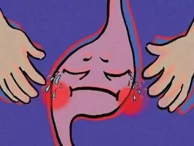
Gastric cancer is one of the most common malignant tumors today. Globally, there are approximately 930,000 new cases of gastric cancer each year, with China accounting for about 42% of these cases. However, due to the often subtle early symptoms of gastric cancer and the lack of regular health check-up habits among many people, most cases are diagnosed at an advanced stage. In the advanced stages of gastric cancer, patients often experience significant suffering, and one of the critical factors severely impacting their quality of life is the development of ascites.
What is gastric cancer ascites?
Ascites refers to the accumulation of fluid in the abdominal cavity beyond physiological limits, meaning an excessive buildup of fluid in the abdomen caused by the shedding of cancer cells into the abdominal cavity.
Clinically, ascites can generally be classified into three main categories: visceral, inflammatory, and neoplastic.
1. Visceral: Cardiac ascites, hepatic ascites, and renal ascites.
2. Inflammatory: Purulent peritonitis, tuberculous peritonitis, and ascites caused by connective tissue diseases.
3. Neoplastic: Gastric cancer, ovarian cancer, colorectal cancer, liver cancer, malignant lymphoma, peritoneal mesothelioma, or metastatic cancerous ascites.
Why does ascites occur in advanced gastric cancer?
In simple terms, gastric cancer ascites occurs when cancer cells shed into the abdominal cavity through various pathways, forming what is referred to as "seeds." These cells often exhibit high biological activity and are a primary cause of peritoneal implantation metastasis and recurrence after surgery.
After peritoneal metastasis, tumor cells damage lymphatic vessels, leading to drainage obstruction. Tumors may invade or compress the thoracic duct, causing impaired lymphatic reflux and leakage of lymph fluid. When lymphatic vessels are blocked or rupture, lymph fluid leaks out, resulting in chylous ascites. Diffuse cancerous infiltration or implantation in the peritoneum increases ascites production. Tumor thrombi block or masses compress the portal vein or hepatic vein, disrupting venous circulation, increasing venous hydrostatic pressure, reducing tissue fluid reabsorption, and causing leakage into the abdominal cavity, leading to ascites. Hypoalbuminemia lowers plasma colloid osmotic pressure, resulting in plasma exudation and ascites formation. In paraneoplastic syndromes, the secretion of ectopic hormones such as antidiuretic hormone and aldosterone increases, causing disturbances in water and electrolyte metabolism and leading to ascites.
Once ascites develops in malignant tumors, common clinical manifestations include abdominal distension and pain, tumor-related pain, difficulty breathing, and lower limb edema. In severe cases, it can lead to renal failure or respiratory failure. These symptoms not only accelerate tumor progression but also significantly impact the patient’s quality of life.

Is Ascites Common in Advanced Gastric Cancer?
How Is It Diagnosed?
Most patients with advanced cancer develop malignant ascites, with a relatively high incidence. Gastrointestinal tumors, liver cancer, and others are particularly prone to this condition. Of course, ascites is also very common in advanced gastric cancer.
The diagnostic process for ascites generally includes the following aspects:
1. Confirming the presence of ascites.
2. Performing diagnostic paracentesis to understand the characteristics of the ascitic fluid.
3. Identifying the cause of ascites based on the patient's medical history, physical signs, and necessary examinations.
How is ascites treated in gastric cancer?
Malignant ascites severely impacts patients' quality of life and is associated with a poor prognosis, with an average survival period of approximately 20 weeks. Therefore, actively treating malignant ascites and improving quality of life are crucial for patients in the terminal stages. The current common treatment options include the following:
1. Initial or Mild Cases, No Specific Treatment Required:
Patients should rest in bed, follow a low-salt diet (daily salt intake limited to 2–4 g), and moderately restrict fluid intake (daily fluid intake around 1–1.5 L).
2. Diuretic Therapy:
Diuretics are generally less effective for malignant ascites. Spironolactone is often the first choice, and it may be combined with furosemide. If the response is inadequate, liver function should be rechecked. If accompanied by hypoalbuminemia (<25 g/L), human albumin infusion can be administered, along with intravenous furosemide (40 mg) or slow intravenous injection of torasemide (10 mg). Electrolytes and urea levels must be monitored during diuretic use to avoid electrolyte imbalances.
3. Paracentesis:
Paracentesis can be performed in malignant ascites patients with symptoms of elevated intra-abdominal pressure, such as nausea, vomiting, abdominal distension, pain, difficulty breathing, or orthopnea. Approximately 90% of patients experience temporary symptom relief, lasting an average of 10.4 days, though repeated procedures are often necessary. It is important to note that repeated large-volume paracentesis carries risks such as reduced effective circulating blood volume, hyponatremia, renal dysfunction, and hypoalbuminemia. Therefore, intravenous albumin or dextran infusions can be used for volume expansion in high-risk patients during fluid drainage.
4. Peritoneal Catheter Drainage:
This is suitable for patients with severe electrolyte imbalances due to paracentesis or those requiring repeated drainage but with contraindications to peritoneovenous shunting (PVS). It rarely causes electrolyte imbalances, poses no risk of cancer cell metastasis or coagulation disorders, and the drainage tube is less prone to blockage.
5. Intraperitoneal Chemotherapy:
Compared to systemic chemotherapy, intraperitoneal perfusion chemotherapy offers significant pharmacokinetic advantages, as it increases local drug concentration in the peritoneal cavity while reducing systemic toxicity.
Drugs that can be used include chemotherapeutic agents (cisplatin, carboplatin, paclitaxel, etc.), sclerosing agents, and biological agents. The response rate generally ranges from 40% to 60%. It is generally recommended that the drug dose should not exceed the intravenous dosage.
6. Hyperthermic Intraperitoneal Chemotherapy (HIPEC):
This involves precise temperature-controlled, circulating perfusion of chemotherapy-containing fluid into the peritoneal cavity, filling it and maintaining it for a specific duration to prevent and treat peritoneal implantation metastasis.
Commonly used drugs include paclitaxel, docetaxel, oxaliplatin, cisplatin, and epirubicin.
The perfusion fluid is primarily normal saline, with a HIPEC volume of 3,000–5,000 ml, a common perfusion flow rate of 300–600 ml/min, and a perfusion duration of 1 hour.
It should be noted that dissolving oxaliplatin or domestic carboplatin in normal saline may cause drug instability. These drugs should be diluted in 5% glucose solution, which may lead to elevated blood glucose during the procedure and requires appropriate management, especially in diabetic patients.
7. Immunotherapy:
Commonly used drugs include interferon, tumor necrosis factor, interleukin, OK-432, Nocardia rubra cell wall skeleton, highly aggregated staphylococcal protein, and Corynebacterium parvum. These are generally administered via intraperitoneal injection, but their efficacy for malignant ascites remains unclear and requires further research for confirmation.
8. Other Treatments:
The management of malignant ascites is a common but complex clinical issue, often requiring comprehensive treatment centered on diuresis, albumin supplementation, and ascites drainage.
During treatment, immune-boosting therapies such as lentinan, thymopentin, or thymalfasin can be administered. Fluid intake should be restricted (1–1.5 L daily), and patients should consume foods high in protein.

Contact Us
Consultation Contact: Director Chen (+86 13884560164)
Medical Address: Gastrointestinal Surgery Department, Digestive Disease Hospital, Gansu Wuwei Academy of Medical and Science (You can directly take Bus No. 13, 15, or 17 to reach the hospital. Alternatively, you can take the hospital's dedicated shuttle bus, which runs every half hour, or a local taxi. Transportation is very convenient.)

