
"Demon Mirror" for All Types of Tumors — PET/CT
"Demon Mirror" for All Types of Tumors — PET/CT
Let me show you the ins and outs of PET/CT
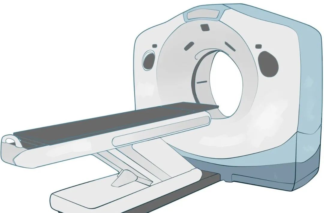
Positron Emission Tomography/Computed Tomography (PET/CT) is an advanced medical imaging technology composed of two core components: PET (Positron Emission Tomography) and CT (Computed Tomography). The PET component excels at revealing the functional and metabolic status of lesions, providing critical diagnostic information for physicians. Meanwhile, the CT component focuses on detailing anatomical structures and density variations, which is equally vital for diagnosis. By integrating the strengths of PET and CT, this technology enhances disease diagnosis in terms of both functional metabolism and anatomical structure, significantly improving sensitivity and accuracy.
The Principle of PET/CT Imaging
Malignant tumor cells are like greedy "predators" within the human body, exhibiting abnormally high metabolic activity as they ruthlessly plunder nutrients. Since glucose serves as one of the primary energy sources for human cells—including tumor cells—malignant tumors exhibit a markedly higher glucose uptake compared to normal tissues.
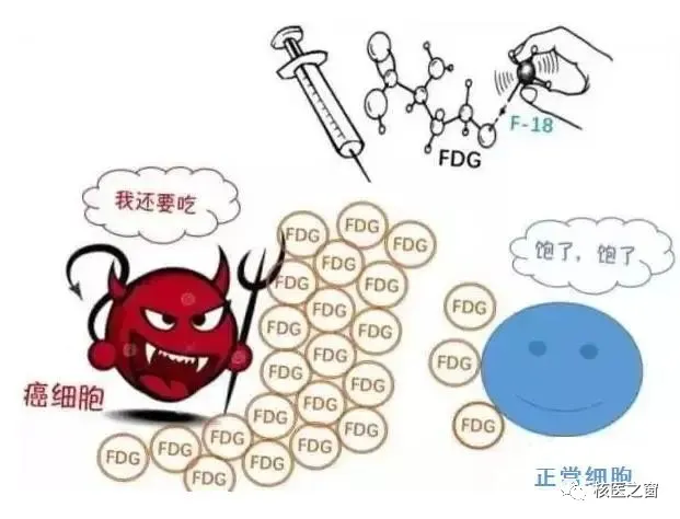
Leveraging the characteristic of malignant tumor cells to intensely uptake glucose, PET/CT technology employs a radiolabeled glucose analog (such as 18F-FDG) as a tracer. After injection into the patient, this tracer accumulates conspicuously in pathological tissues like tumors, creating distinct bright spots on PET/CT images. The degree of accumulation can be quantitatively assessed through the Maximum Standardized Uptake Value (SUVmax). As a crucial semi-quantitative metric in oncological PET/CT diagnostics, SUVmax provides physicians with an objective basis for evaluating tumor activity and metabolic status.
Why Can PET/CT Detect Tumors?
Before undergoing a PET/CT examination, patients are injected with a tumor-targeting molecular probe drug called 18F-FDG. This radiopharmaceutical travels through the body's physiological transport and metabolic pathways, selectively accumulating within tumor cells. Inside tumor cells, 18F-FDG is converted into 6-phospho-deoxyglucose (6-PDG) by the enzyme hexokinase—a key glycolytic enzyme. This transformation traps 18F-FDG inside the cells, as the phosphorylated form cannot easily exit. Notably, the metabolic activity of tumor cells directly correlates with their ability to uptake and retain 18F-FDG: the more metabolically active the tumor cells, the greater their accumulation of the tracer.
By leveraging PET/CT technology, physicians can precisely localize tumor cells, determine their size, and assess their morphological characteristics. This capability not only provides comprehensive diagnostic information but also significantly enhances the accuracy and reliability of tumor detection.
Which Diseases Can Be Diagnosed Using PET/CT?
The primary applications of PET/CT in oncology include:
1. Early Diagnosis of Tumors
By enabling early detection and timely intervention, many tumor cases can potentially be cured.
2. Preoperative Assessment
Through comprehensive evaluation of the disease, we can develop personalized treatment plans for patients. For confirmed in situ cancers, surgery is often the preferred treatment; whereas in cases of extensive metastasis, surgical intervention may be unnecessary, and radiotherapy or chemotherapy can be directly employed. This assessment process is crucial for guiding clinicians in selecting the most appropriate therapeutic strategy.
3. Differential Diagnosis of Benign vs. Malignant Tumors
When it is challenging to distinguish between benign and malignant tumors, PET/CT technology can provide precise diagnostic differentiation.
4. Locating the Primary Tumor
For many patients with elevated tumor markers but an unclear tumor location, PET/CT's whole-body imaging can accurately track and pinpoint the primary lesion.
5. Biological Target Volume (BTV) Delineation and Surgical Resection Planning
By integrating PET functional imaging, PET/CT enables a metabolic-level understanding of tumor characteristics. Based on BTV mapping from PET/CT, we can precisely define radiation therapy targets and surgical resection margins, providing robust support for personalized treatment planning.
6. Guiding Tumor Biopsy for Surgeons
While pathological examination remains the "gold standard" for tumor diagnosis, clinicians often face challenges in identifying optimal biopsy sites. PET/CT imaging—by highlighting biological target zones—provides critical guidance for tissue sampling, significantly improving diagnostic accuracy and efficacy.
7. Some highly suspected patients and high-risk populations
PET/CT is recommended for individuals with premalignant lesions, a family history of cancer, those engaged in high-risk occupations over prolonged periods, and people who have experienced unexplained progressive weight loss within a short timeframe.
8. Assessment of Cardiac Myocardial Cells
PET/CT is regarded as the "gold standard" in evaluating myocardial cell viability. Before performing bypass surgery, it is crucial to assess the viability of the patient's myocardial cells, making nuclear medicine techniques such as myocardial perfusion imaging and myocardial metabolic imaging the optimal choices. Additionally, PET/CT is widely used to evaluate the outcomes of myocardial bypass surgery, providing physicians with precise postoperative feedback.
9. Application in Neurological Diseases
PET/CT plays a significant role in brain function assessment. It is not only used for the diagnosis of dementia but also enables accurate identification of gliomas, precise clinical staging, and effective prediction of tumor recurrence.
Common Questions About PET/CT
1. What are the advantages of PET/CT?
As an advanced medical imaging technology, PET/CT can detect life-threatening diseases such as tumors at an early stage. In the initial phases of tumor development, through detailed metabolic molecular imaging, PET/CT can sensitively identify traces of malignant tumors. As a crucial tool for early screening in high-risk populations, PET/CT not only precisely locates lesions anatomically but also provides a metabolic-level analysis. This dual capability strongly supports the goals of early detection, early diagnosis, and early treatment.
2. Do I need other tests after a PET/CT scan?
While PET/CT is undoubtedly one of the most advanced medical imaging technologies available today, it does have certain limitations. In tumor detection, particularly when tumors are very small (measuring less than 5-8 mm in size), PET/CT's resolution may not be sufficient, posing a risk of missed diagnosis. Moreover, the field of medical imaging encompasses various modalities, including CT, MRI, ultrasound, and gastrointestinal endoscopy, each with its own unique strengths and specific applications. PET/CT is just one component of the diagnostic toolkit and cannot completely replace other examination methods. Therefore, the choice of specific follow-up tests should be determined based on your physician's professional recommendation and your individual clinical condition, ensuring the most accurate and comprehensive diagnostic evaluation.
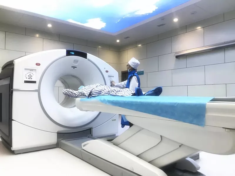
The PET-CT Center at Gansu Wuwei Medical Science Academy Cancer Hospital has introduced the GE Discovery 710 Clarity Super Iterative PET-CT from the United States, which is currently the most advanced 256-ring LBS crystal PET/128-slice spiral CT in China. This equipment is the fourth PET-CT in Gansu Province and the only one in prefecture-level hospitals across the province. As a super iterative device launched by GE that leads the global trend, it offers higher sensitivity and specificity compared to conventional PET/CT systems. With its exclusive 1.8mm high spatial resolution, it significantly enhances the detection capability for tiny lesions while substantially reducing whole-body scan time and radiation exposure for patients, delivering optimal diagnostic results - earning it the reputation as the crown jewel of modern medical high technology. The center features first-class radiopharmaceutical preparation facilities, equipped with a medical cyclotron system capable of producing various disease-specific diagnostic tracers including 11C, 13N, and 18F. The purity of the pharmaceuticals produced exceeds national standards, reaching world-class levels to ensure accurate diagnoses for patients. Our PET-CT Center has become the only facility in Gansu Province capable of performing both 11C and 18F imaging examinations.
Contact us
Consultation phone: Director Li Ke ( +86 13689357660)
Medical address: PET-CT Center, Heavy Ion District, Wuwei Academy of Medical and Science