
Medical Physicists: Crafting Your Personalized Radiotherapy Plan
Medical Physicists: Crafting Your Personalized Radiotherapy Plan
When undergoing radiotherapy, patients often hear the term "radiotherapy plan." So, what exactly is a radiotherapy plan, and how is it customized for each individual?
This article will walk you through the process of creating a radiotherapy plan and the important role that medical physicists play in that process.
1. Why is “one person, one plan” necessary?
Radiotherapy, short for radiation therapy, is a treatment modality that uses high‑energy radiation to "precisely eliminate" tumor cells. It is like a "precision sniper attack" against the tumor – maximizing the destruction of the "enemy" (tumor cells) while doing its utmost to protect the "civilians" (surrounding critical organs).
Because each patient's tumor condition and physical status are unique, there can never be a "one‑size‑fits‑all template." The reason radiotherapy plans must be "one plan per patient" is based on the following three key determinants:
1. The Tumor's "Identity Information": Size, Location, and Shape
This is the most core determinant. Just as reconnaissance is needed before a battle, physicians need to accurately identify the following using imaging such as CT, MRI, or even PET‑CT:
Where is the enemy? Is the tumor located in the lung, prostate, or nasopharynx? The depth and location affect the path the radiation beam must travel and the "minefields" (critical organs) it must avoid.
How big is the enemy, and what does it look like? Is the tumor a regular sphere or an irregular "crab‑like" shape? The plan must precisely delineate its three‑dimensional contour (known medically as the "target volume") to ensure that the radiation beam completely covers the tumor without leaving any gaps.

Figure 1. Radiation therapy planning: comprehensive tumor analysis
2. The Patient's "Individual Map": Body Structure and Adjacent Organs
Every person has subtle differences in their internal anatomical structure. The radiotherapy plan highlights the "key protected structures" around the tumor, such as:
For head and neck tumors: the adjacent spinal cord, brainstem, eyes, etc.
For thoracic tumors: the adjacent heart, lungs, esophagus, etc.
For prostate tumors: the adjacent bladder, rectum, etc.
The dose distribution in the plan must be as precise as a detailed map, ensuring that the high‑dose region exactly matches the shape of the tumor, while allowing the surrounding normal tissues to stay out of the high‑dose area as much as possible.

Figure 2. Schematic diagram of some critical organs around tumors at different locations.
3. The Patient's "Baseline Conditions": Physical Status and Treatment History
Factors such as the patient's age, overall health status, presence of other comorbid conditions (e.g., heart disease, pulmonary fibrosis), and prior treatment history (e.g., surgery or radiotherapy) all significantly influence the radiotherapy plan.
When developing the plan, the goal is to minimize treatment‑related risks and improve overall tolerance, while ensuring therapeutic efficacy.
2. The core design ideas of physicists: seeking optimal solutions amid contradictions
The specific design of a radiotherapy plan is carried out by a key professional: the medical physicist. Their work is truly an art of "seeking the best balance among multiple conflicting objectives." The core approach is as follows:
Step 1: Define the Goals – "Where to Hit, Where to Spare"
The radiation oncologist delineates the tumor area to be irradiated (the target volume) and identifies the normal organs to be protected, setting dose objectives for each region:
For the tumor: It must receive the "prescription dose" sufficient to kill cancer cells, and the dose distribution should be uniform.
For normal organs: The dose must remain below the organ‑specific tolerance limits to avoid the risk of damage.
Step 2: Choose the Weapon and Path – "What to Use and Where to Deliver From"
Based on the tumor location, the physicist selects the most suitable radiotherapy technique. They must design the "delivery path": deciding from which angles to deliver the radiation and how to dynamically adjust the intensity of the beam from each angle. This process relies on powerful treatment planning systems to perform extensive calculations. The sole objective is to make the high‑dose region tightly "hug" the tumor while dropping off sharply like a cliff in the surrounding normal tissues.

Figure 3. Comparison of two technologies, IMRT (left) and VMAT (right)
Step 3: Iterative Optimization and Validation – "Fine‑Tuning to Ensure Flawless Delivery"
No plan is perfect on the first try. The physicist repeatedly adjusts parameters, performing dozens or even hundreds of simulations on the computer, comparing the pros and cons of different approaches. Using professional tools such as dose‑volume histograms for evaluation, they continue until they find a plan that achieves the best possible balance between destroying the tumor and protecting normal tissues.
Once the plan is finalized, treatment cannot begin immediately. The physicist then uses specialized equipment or phantoms to perform dose verification, ensuring that the perfect plan designed on the computer can be executed accurately and precisely by the treatment machine. This serves as the final safety checkpoint before treatment.
3. As a patient, how can you cooperate in making a good plan?
A successful radiotherapy plan relies on the joint efforts of both the medical team and the patient. As a patient, you can contribute in the following ways:
1. Communicate Fully and Provide Complete Information
During your consultation, provide your physician with detailed information about your full medical history, past treatments (especially surgeries and radiotherapy), and all current symptoms. This information is critical for the physician to assess your condition and determine safe dose levels.

Figure 4. Fully communicate and provide complete medical history information
2. Cooperate Fully with "Simulation" and "Immobilization"
The first step of radiotherapy is the "simulation CT scan." It is essential to maintain the exact position required by your physician and to remain completely still throughout the scan. This position will be your standard treatment position for every future session. For areas requiring immobilization (such as the head or neck), you may feel slight pressure when a body mold or face mask is being made. Please cooperate as much as possible – good immobilization is the foundation of precise treatment.
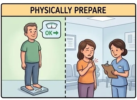
Figure 5. Schematic diagram of patient positioning
3. Prepare Your Body Before Treatment
- Maintain stable body weight: Significant weight gain or loss changes your body contour and internal anatomy, potentially making the originally meticulously designed plan "inaccurate."
- Manage relevant symptoms: If you have symptoms that affect your ability to maintain the treatment position, such as pain or difficulty breathing, inform your medical team promptly. They will help alleviate these symptoms to ensure consistent positioning for each treatment session.

Figure 6. Symptoms related to weight control and management
4. Perform the "Standard Actions" During Treatment
Each time you lie on the treatment couch, try to precisely reproduce the position from your simulation session. Breathe calmly, relax your whole body, and follow the instructions of the radiation therapists. Your good cooperation is key to the flawless execution of the treatment plan.
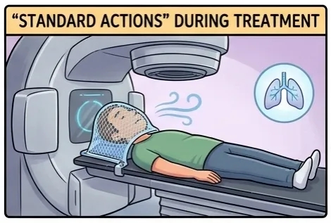
Figure 7. The patient is fixed in a fixed position for treatment.
5. Actively Report Treatment Reactions
During the course of treatment, promptly report any discomfort you experience (such as skin reactions, pain on swallowing, fatigue, etc.) to your physician. This helps the doctor determine whether the reactions are within expected limits and allows for supportive care or plan adjustments if necessary.

Figure 8. Positive feedback on treatment response
Behind the "one plan per patient" approach in radiotherapy lies the relentless pursuit of "precision" and "individualization" in modern oncology. It is not merely a set of cold technical parameters, but rather a personalized, compassionate life‑safeguarding plan meticulously crafted for you by professional medical staff. Understand it, trust it, and actively cooperate with it – and you will have the most reliable ally in this precise battle against the disease.
Author: Meng Wanbin
Initial Review: Liu Qiong
Secondary Review: Guo Yishan
Secondary Review:Cai Qinghua
Lanzhou Campus of Gansu Wuwei Cancer Hospital – Consultation Hotline:
+86 166 0931 3666 – Director Li
+86 176 9325 0603 – Ms. Yan