A New Precision Option in Cancer Care: Clinical Cases of Carbon‑Ion Therapy
A New Precision Option in Cancer Care: Clinical Cases of Carbon‑Ion Therapy
I. Clinical Case Presentations
Case 1: Patient Zhang – Malignant Pancreatic Tumor
1. Brief Medical History:
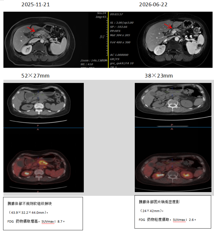
Patient Zhang, female, 63 years old, was admitted with "6 months post‑carbon‑ion radiotherapy for pancreatic body cancer."
In October 2025, she underwent a whole‑abdomen CT due to "intermittent upper abdominal distension." The results showed a weakly enhancing mass in the pancreatic body, suggestive of pancreatic cancer, with involvement of the splenic artery and vein. Endoscopic ultrasound‑guided fine‑needle aspiration biopsy confirmed pancreatic ductal adenocarcinoma, well‑differentiated.
PET‑CT revealed an irregular soft tissue mass in the pancreatic body, with ill‑defined margins and indistinct demarcation from the adjacent gastric wall, with increased metabolic activity — consistent with pancreatic cancer on PET‑CT.
From December 3, 2025, she received curative carbon‑ion radiotherapy for pancreatic cancer at a dose of PTV 39.6 Gy(RBE) in 9 fractions, with a boost of PTV 17.6 Gy(RBE) in 4 fractions, along with concurrent intravenous gemcitabine chemotherapy. Mild gastrointestinal adverse reactions were observed during the treatment course.
From January 12, 2026, to June 26, 2026, she completed 12 cycles of systemic intravenous chemotherapy with the NALIRIFOX regimen.
2. Imaging Comparison
Case 2: Patient Wang – Malignant Pancreatic Tumor
1. Brief Medical History:
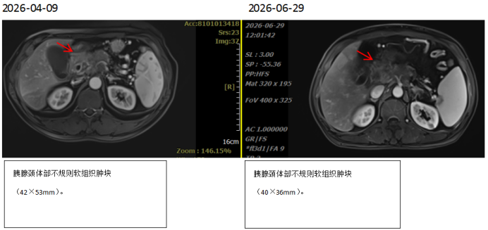
Patient Wang, male, 60 years old, was admitted with "1 month post‑carbon‑ion therapy for pancreatic neck‑body cancer." In February 2026, he underwent an abdominal CT due to "upper abdominal pain radiating to the back," which revealed a soft tissue mass above the pancreatic head, measuring approximately 54 × 40 mm in maximum cross‑section, with involvement of the celiac trunk and splenic vein. Endoscopic ultrasound‑guided pancreatic biopsy suggested adenocarcinoma.
PET‑CT showed a soft tissue density mass in the pancreatic neck‑body region, measuring approximately 38 mm × 43 mm × 28 mm, with ill‑defined borders and increased FDG uptake (SUVmax: 7.2), consistent with pancreatic cancer on PET‑CT.
From April 10, 2026, he received carbon‑ion radiotherapy for the pancreatic lesion with the following prescription doses: PTV40: 40 Gy(RBE) in 10 fractions, PTV48: 48 Gy(RBE) in 2 fractions, PTV56: 56 Gy(RBE) in 2 fractions, with concurrent intravenous gemcitabine chemotherapy. After completion of treatment, the upper abdominal and back pain resolved completely.
From May 14 to June 6, 2026, he completed 2 cycles of systemic intravenous chemotherapy with the NALIRIFOX regimen.
2. Imaging Comparison:
II. Summary
With its advantages of precision, high biological effectiveness, and low damage to surrounding tissues, carbon‑ion therapy has emerged as a significant breakthrough in the treatment of refractory abdominopelvic tumors, including pancreatic cancer and duodenal lymphoma. The Department of Radiation Oncology III at Gansu Wuwei Cancer Hospital, supported by a specialized team, advanced equipment, and extensive clinical experience, has achieved remarkable outcomes in carbon‑ion therapy. These results not only offer renewed hope to patients already treated but also open new therapeutic pathways for more patients with locally advanced pancreatic cancer through ongoing clinical research. If you or someone close to you meets the eligibility criteria for our clinical study, please contact us through the methods provided to learn more and join us in this journey of hope against cancer.