
Patient, female, 61 years old, pancreatic malignancy
Patient: Yang XX, female, 61 years old
Chief Complaint: Hospitalized due to "confirmed pancreatic cancer for 1 year."
[Clinical Summary]
- Imaging Studies: Abdominal MRI: A mass in the body and tail of the pancreas, invading the celiac trunk, superior mesenteric artery, and splenic vein, accompanied by pancreatic portal hypertension. The lesion shows poorly defined borders with the left adrenal gland and gastric wall.
- Pathological Diagnosis: Pancreatic mass biopsy confirmed adenocarcinoma.
[Clinical Diagnosis]
Malignant pancreatic tumor (body and tail, adenocarcinoma, cT4N2M0 Stage III, KPS score: 90).
[Treatment Plan]
- Carbon-ion radiotherapy: Total dose 58.5 Gy (RBE)/14 fractions.
- Concurrent chemotherapy: NALIRIFOX regimen (liposomal irinotecan + oxaliplatin + leucovorin + 5-fluorouracil).
[Treatment Response]
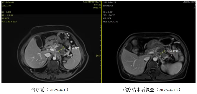
Left image: Before treatment; Right image: One month after treatment
This case demonstrates the significant efficacy of heavy-ion radiotherapy in the treatment of locally advanced and metastatic pancreatic cancer. Through personalized treatment, the patient exhibited a marked reduction in tumor marker levels and significant imaging improvement.
Moreover, carbon-ion therapy not only demonstrates remarkable tumor suppression during treatment but also exhibits sustained biological advantages post-treatment. This effect allows the tumor to continue shrinking for 3-6 months after therapy, reaching its peak response at 6 months. Due to carbon ions inducing DNA double-strand breaks, their tumoricidal effect is more thorough, effectively reducing recurrence rates and improving local control. This approach provides patients with longer progression-free survival and enhanced quality of life.For patients with locally advanced or inoperable pancreatic cancer, this technology undoubtedly opens a promising therapeutic pathway.