Patient, male, 57 years old, pancreatic malignancy
Patient, male, 57 years old, pancreatic malignancy.
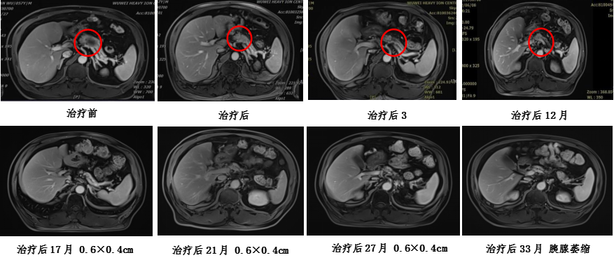
Medical history: The patient was found to have a tumor marker CA199 greater than 1000 during a physical examination on April 27, 2021. He then sought medical attention at Gansu Provincial People's Hospital on May 7th, where a full abdominal MRI showed a mass in the body of the pancreas (approximately 6.2cm × 3.5cm in size), suggestive of pancreatic cancer. Tumor marker testing indicated CA199 greater than 1200. PET-CT scan revealed low-density nodules with increased metabolism in the body of the pancreas, indicating a tendency towards pancreatic cancer. The patient visited our department in May 2021, and abdominal MR imaging showed a mass in the pancreatic neck with unclear local boundaries with the splenic vessels. Ultrasound-guided endoscopic biopsy of the pancreatic lesion was performed, and pathological examination revealed moderately differentiated ductal adenocarcinoma.
Diagnosis: Malignant pancreatic tumor (moderately differentiated ductal adenocarcinoma in the body of the pancreas, cT4N1M0 stage III, KPS score: 80)
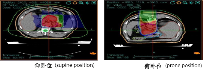
Treatment plan: Carbon ion radiotherapy for pancreatic cancer, supine position with three-field irradiation, total dose: 41.4GyE, 4.6GyE/fraction; prone position PTVboost for local boost dose of 18.4GyE/4 fractions, 4.6GyE/fraction, total prescription dose: PTV 59.8GyE/13 fractions. During treatment, combined with intravenous injection of Nimotuzumab 400mg on day 1 weekly + Gemcitabine 2.0g on days 1 and 8 for chemotherapy. The treatment process proceeded smoothly without significant adverse reactions. Follow-up abdominal MRI after treatment showed a reduction in the pancreatic lesion, with treatment response assessed as partial response (PR).
Carbon ion treatment plan dose distribution map and efficacy evaluation: