
Carbon ion therapy for lung cancer: Two fractionation modes in lymph node drainage area
Clinical report of two dose fractionation modes using carbon ion beam therapy in the lymph node drainage area for lung cancer
Pan Xin1, Zhang Yihe1, Li Xiaojun1, Ma Tong1, Wang Xin1, Yang Yuling1, Chai Hongyu1, Qin Tianyan2, Lyu Caixia2, Li Pengqing2, Ye Yancheng1, Zhang Yanshan1
1 Department of Radiation Oncology, Heavy Ion Center, Wuwei Cancer Hospital of Gansu Province, Wuwei 733000, China; 2 Registration / Follow ‐ up Center, Heavy Ion Center, Wuwei Cancer Hospital of Gansu Province, Wuwei 733000, China
Corresponding author: Zhang Yanshan, Email: 13830510999 @163.com
【 Abstract 】 Objective To compare the adverse reactions, efficacy and survival rate of carbon ion beam irradiation in the elective lymph node (ENI) drainage area of locally advanced non ‑ small cell lung cancer (LA‑NSCLC) with relative biological effect (RBE) dose of 48 Gy using 16 and 12 fractions. Methods A total of 72 patients with pathologically confirmed LA ‑ NSCLC admitted to Wuwei Heavy Ion Center of Gansu Wuwei Tumor Hospital from June 2020 to December 2021 were enrolled and simple randomly divided into groups A and B, with 36 patients in each group. Patients in groups A and B were treated with carbon ion beam irradiation to the lymph node drainage area with 48 Gy (RBE) using 16 and 12 fractions. The acute and chronic adverse reactions, efficacy and survival rate were observed. The survival curve was drawn by Kaplan‑Meier method. Difference test was conducted by log‑rank test. Results The median follow‑up time was 13.9 (8.8 ‑ 15.7) months in group A and 14.6 (6.3 ‑ 15.9) months in group B. Sixteen (44.4%) patients were effectively treated in group A and 9 (25%) patients in group B. Thirty ‑ four (94.4%) cases achieved disease control in group A and 30 (83.3%) cases in group B. Statistical analysis showed that the overall survival rate in group B was similar to that in group A (χ2=1.192, P=0.275). Comparison of planning parameters between two groups showed CTV volume, Dmean, V5 Gy(RBE), V20 Gy(RBE) and V30 Gy(RBE) of the affected lung, cardiac V20 Gy(RBE), V30 Gy(RBE) and Dmean, esophageal V30 Gy(RBE), V50 Gy(RBE), Dmax and Dmean, Dmax of the trachea and spinal cord had no significant difference (all P>0.05). No grade 3 or 4 adverse reactions occurred in the enrolled patients during treatment and follow‑up. No statistical differences were observed in the acute radiation skin reaction (χ2=5.134, P=0.077), radiation esophagitis (χ2=1.984, P=0.371), and advanced radiation pneumonia (χ2=6.185, P=0.103) between two groups. Conclusions The two dose fractionation modes of carbon ion therapy system are equally safe in the mediastinal lymphatic drainage area of LA ‑ NSCLC, and the adverse reactions are controllable. The long ‑ term efficacy still needs further observation.
【Key words】 Radiotherapy, carbon ion; Carcinoma, non‑small cell lung; Elective lymph node drainage area radiotherapy; Adverse reactions; Short‑term efficacy
Fund programs: Key Science and Technology R&D Plan of Gansu Province ‑ Construction of Heavy Ion Treatment Center for Social Development ( 19YF3FH001); Natural Science Foundation of Gansu Province ‑ 2021 Innovation Base and Talents Program of Science and Technology Department of Gansu Province (21JR7RH896)
Currently, the incidence and mortality rates of lung cancer remain high worldwide. One of the main reasons for the poor prognosis of lung cancer patients is that the disease is often detected at an advanced stage, when surgical intervention is no longer feasible. For patients with stage I non-small cell lung cancer (NSCLC), surgical treatment can achieve a 10-year survival rate of up to 92% [1]. Therefore, early detection of lung cancer, along with accurate staging and selection of appropriate treatment strategies, is of great significance for improving patient outcomes.
Verhagen et al. [2] reported that even after curative surgery, the postoperative recurrence rate for stage I NSCLC patients remains as high as 25%–50%, with lymph node metastasis being one of the primary factors contributing to recurrence. In the context of surgical treatment for lung cancer, lobectomy combined with lymph node dissection is the standard recommended approach. For instance, both the 2020 National Comprehensive Cancer Network (NCCN) guidelines and the 2020 Chinese Society of Clinical Oncology (CSCO) guidelines recommend hilar and mediastinal lymph node dissection even for T1-stage lung cancer. Before computed tomography (CT) was used for radiotherapy planning, elective node irradiation (ENI) was the standard radiotherapy strategy for NSCLC. Its advantage lay in reducing the risk of regional lymph node failure [3]. However, because ENI targets not only the primary tumor and metastatic lymph nodes but also clinically negative lymph node drainage areas, it inevitably exposes excessive normal tissues—such as the lungs, esophagus, and heart—to radiation. This often leads to severe complications like radiation pneumonitis and esophagitis, which in turn limit the ability to safely escalate the radiation dose to the primary tumor. The unique physical and biological advantages of carbon ion beam therapy now make it possible to revisit the ENI strategy with improved precision and reduced toxicity.
This study utilized a carbon ion therapy system to treat locally advanced non-small cell lung cancer (LA-NSCLC). The lymph node drainage areas were irradiated with carbon ion beams at doses of 48 Gy (RBE) in 16 fractions and 48 Gy (RBE) in 12 fractions, respectively. The aim was to observe acute and chronic adverse reactions, treatment efficacy, and survival rates, thereby providing reference for the formulation and optimization of carbon ion radiotherapy plans for LA-NSCLC.
Data and Methods
1. Patient data: A total of 72 patients with pathologically confirmed LA-NSCLC admitted to the Heavy Ion Center of Gansu Wuwei Cancer Hospital between June 2020 and December 2021 were selected. Staging was performed using cranial MRI, chest CT, and PET-CT according to the 8th edition of the American Joint Committee on Cancer (AJCC) staging system. Patients with stages T1-3N1-2M0 and T3-4N0-1M0 were randomly divided into group A and group B, with 36 patients in each group. This study was approved by the Ethics Committee of Gansu Wuwei Cancer Hospital (Approval No. 2021-Ethical Review-05).
2. Carbon ion therapy
(1) Patients were treated in the supine position, lying flat on the positioning couch with a fixed pillow under the head. Both arms were raised and crossed above the head, with elbows fully extended as much as possible. The body position was immobilized using a thoracic thermoplastic mask and a vacuum cushion. CT scanning included both plain and enhanced sequences, along with 4DCT imaging. The slice thickness and interval were both set at 3 mm, and the scanning range extended from the patient’s cricothyroid membrane to the lower edge of the first lumbar vertebra.
(2) Target volume delineation: Target areas were contoured using standardized protocols (with fusion of MRI or PET-CT images when necessary). GTV referred to the gross tumor volume visible on imaging (referencing enhanced CT, MRI, or PET-CT). GTVnd denoted metastatic mediastinal lymph nodes, defined as those with a short-axis diameter ≥1 cm and/or PET-CT positivity and/or pathological confirmation via endoscopic ultrasound. CTV encompassed GTV plus a 0.5 cm margin around GTVnd, along with elective nodal irradiation coverage (including involved fields of metastatic nodes and prophylactic regions based on nodal metastasis risk, typically covering lymph node groups Ⅱ, Ⅳ, Ⅴ, and Ⅶ). ITV represented the range of CTV motion observed on 4DCT. PTV was generated by expanding ITV by 3–5 mm, with adjustments made near organs at risk. Carbon ion treatment plans were developed using the Ci-Plan carbon ion treatment planning system (version 1.0). Prescription doses: For Group A, the elective nodal irradiation dose was 48 Gy (RBE) in 16 fractions; for Group B, it was 48 Gy (RBE) in 12 fractions. The total dose to the primary tumor site was 72 Gy (RBE) for both groups.
3. Follow-up: Patients were followed up via outpatient clinics for over six months, starting from the first day after radiotherapy completion. The first two follow-ups were conducted monthly, followed by quarterly visits until February 28, 2022.
4. Efficacy evaluation and adverse reaction assessment: Patients included in the study were clinically observed. Acute adverse reactions were evaluated using the Common Terminology Criteria for Adverse Events (CTCAE) v5.0, while late adverse reactions were assessed based on the Radiation Therapy Oncology Group (RTOG) criteria. Treatment efficacy for the two dose fractionation regimens was determined according to the Response Evaluation Criteria in Solid Tumors (RECIST 1.1).
Local control rate = (complete response + partial response + stable disease) / number of evaluable cases × 100%;
Time to progression (TTP) was defined as the duration from the start of radiotherapy to local recurrence;
Overall survival (OS) was defined as the duration from the start of radiotherapy to death from any cause or the last follow-up.
5. Statistical analysis: Data were processed and analyzed using SPSS 20.0 software. Measurement data are expressed as mean ± standard deviation (x̄ ± s), while enumeration data are presented as percentages. The t-test and chi-square test were employed to analyze countable data, survival periods were assessed using the Kaplan-Meier method, and short-term adverse reactions were compared with the chi-square test. A P-value <0.05 was considered statistically significant.
Results
1. General data: A total of 72 pathologically confirmed NSCLC patients were included in the study. In both Group A and Group B, the male-to-female ratio was 26:10 (72.2%:27.8%), with median ages of 62.5 years (range: 55–70) and 62 years (range: 51.5–68.5), respectively. Regarding pathological types, squamous cell carcinoma (17 cases, 47.2%) and adenocarcinoma (17 cases, 47.2%) accounted for the majority in Group A, while Group B had 17 cases (47.2%) of squamous cell carcinoma and 15 cases (41.7%) of adenocarcinoma. Based on the Chinese Society of Clinical Oncology (CSCO) guidelines for NSCLC treatment principles and the patients' actual conditions, 22 patients (61.1%) in Group A received concurrent chemotherapy, compared to 31 patients (86.1%) in Group B. Comparison of baseline clinical data showed a statistically significant difference in Karnofsky Performance Status (KPS) scores between the two groups (χ²=28.39, P<0.001), while no significant differences were observed in other indicators. The general characteristics of the patients are summarized in Table 1.
2. Treatment outcomes: According to the study design, both groups received a total dose of 48 Gy (RBE) to the elective nodal irradiation area, but with different fractionation schemes: 3 Gy (RBE) × 16 fractions for Group A and 4 Gy (RBE) × 12 fractions for Group B. The plan design is illustrated in Figure 1. To evaluate the dose exposure to organs at risk (OARs), relevant parameters were statistically analyzed. The results indicated no statistically significant differences in the following metrics: GTV volume, CTV volume, mean dose (Dmean) to the ipsilateral lung, V5 Gy (RBE), V20 Gy (RBE), V30 Gy (RBE) of the ipsilateral lung; V20 Gy (RBE), V30 Gy (RBE), and Dmean of the heart; V30 Gy (RBE), V50 Gy (RBE), maximum dose (Dmax), and Dmean of the esophagus; Dmax of the trachea; and Dmax of the spinal cord. Detailed data are presented in Table 2.
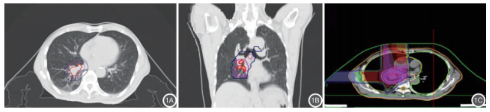
Figure 1 Example of elective nodal irradiation planning design for mediastinal lymph node drainage areas in patients with locally advanced non-small cell lung cancer treated with carbon ion radiotherapy (1A, 1B: Target volume delineation; 1C: Carbon ion plan dose distribution cloud diagram)
Table 1 Baseline clinical characteristics of 72 patients with locally advanced non-small cell lung cancer treated with carbon ion radiotherapy [n (%) or M (range)]
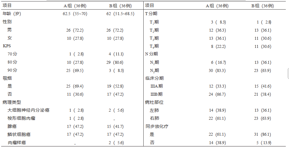
Note: Group A received a radiotherapy dose of 3 Gy (RBE) × 16 fractions; Group B received a radiotherapy dose of 4 Gy (RBE) × 12 fractions; KPS = Karnofsky Performance Status; RBE = Relative Biological Effectiveness
Table 2 Dosimetric parameters of organs at risk for 72 patients with locally advanced non-small cell lung cancer treated with carbon ion radiotherapy under different fractionation schemes [M (range)]
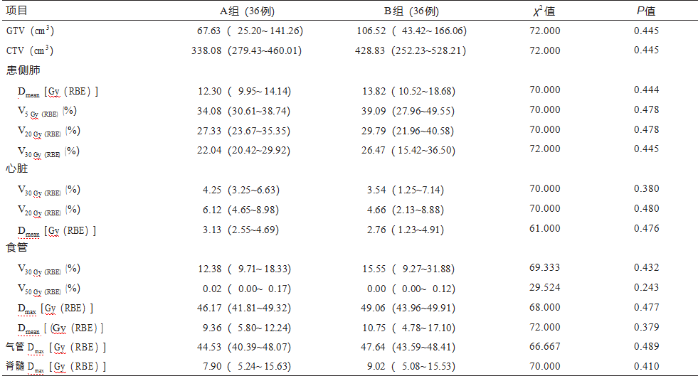
Note: Group A received a radiotherapy dose of 3 Gy (RBE) × 16 fractions; Group B received a radiotherapy dose of 4 Gy (RBE) × 12 fractions; RBE = Relative Biological Effectiveness.
Patients in both Group A and Group B completed the prescribed carbon ion therapy as planned. No Grade 3 or 4 adverse reactions were observed during treatment or throughout the follow-up period. Although dosimetric comparisons showed no significant differences in organ-at-risk parameters between the two groups, some patients received concurrent chemotherapy during carbon ion therapy due to variations in their clinical conditions. Further analysis of early and late adverse reactions revealed no statistically significant differences between the two groups in terms of acute radiation dermatitis (χ²=5.134, P=0.077), radiation esophagitis (χ²=1.984, P=0.371), or late radiation pneumonitis (χ²=6.185, P=0.103). Detailed data are presented in Table 3.
Table 3 Comparison of adverse reactions in 72 patients with locally advanced non-small cell lung cancer treated with carbon ion radiotherapy [n (%)]
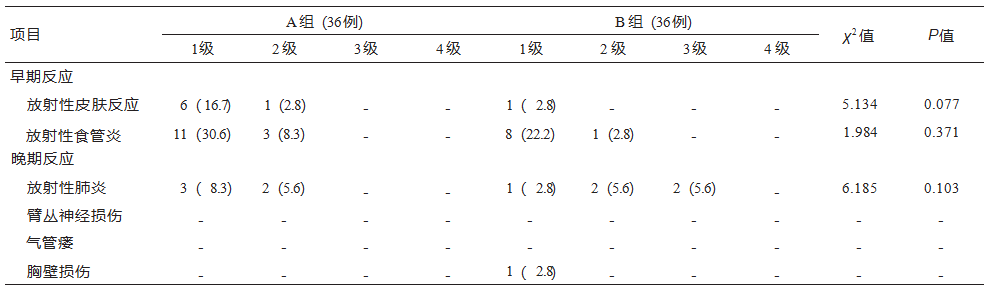
Note: Group A received a radiotherapy dose of 3 Gy (RBE) × 16 fractions; Group B received a radiotherapy dose of 4 Gy (RBE) × 12 fractions.
3. Patient prognosis
(1) Comparison of short-term efficacy between the two groups: The follow-up period for all patients in this study ended on February 28, 2022. All enrolled patients were effectively followed up with valid follow-up data obtained. The median follow-up time was 13.9 months (range: 8.8-15.7) for Group A and 14.6 months (range: 6.3-15.9) for Group B. During follow-up, 1 case (2.8%) of in-field recurrence occurred in Group A, while 2 cases (5.6%) occurred in Group B. All other disease progression cases were due to distant metastasis or new metastatic lesions.
Specifically:
• Treatment response was observed in 16 cases (44.4%) in Group A and 9 cases (25.0%) in Group B, with no statistically significant difference (χ²=3.003, P=0.083).
• Disease control was achieved in 34 cases (94.4%) in Group A and 30 cases (83.3%) in Group B, with no statistically significant difference (χ²=2.250, P=0.134).
(2) Comparison of survival outcomes between the two groups:
The Kaplan-Meier method was used to plot survival curves, analyzing local control rates and overall survival during the follow-up period for both groups. The log-rank test was applied for difference testing. The analysis showed that Group B had similar local control rates (χ²=0.517, P=0.472) and overall survival rates (χ²=1.192, P=0.275) compared to Group A. The mean LRFS periods were 20.8 months (95% CI: 19.63–20.06) for Group A and 17.1 months (95% CI: 15.39–18.79) for Group B. The mean OS periods were 21.1 months (95% CI: 20.01–22.29) for Group A and 16.8 months (95% CI: 15.27–18.42) for Group B. No statistically significant differences were observed (P>0.05), indicating similar overall survival between the two groups by the end of follow-up, as shown in Figure 2.
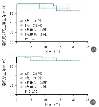
Figure 2 Comparison of local progression-free survival (2A) and overall survival (2B) between the two groups of patients with locally advanced non-small cell lung cancer treated with carbon ion radiotherapy (n=72) [Radiation doses: Group A: 3 Gy (RBE) × 16 fractions; Group B: 4 Gy (RBE) × 12 fractions; RBE = Relative Biological Effectiveness]
Discussion
Regional lymph node metastasis is a major prognostic factor in NSCLC patients. During pulmonary resection, it is recommended to remove both intrapulmonary and mediastinal lymph nodes simultaneously. Despite intended radical treatment, 25% to 50% of early-stage lung cancer patients still experience recurrence during follow-up, suggesting that early lymphatic dissemination of isolated tumor cells and micrometastases may influence patient outcomes [4].
ENI (Elective Nodal Irradiation) encompasses the primary tumor, metastatic lymph nodes, and clinically negative lymph node drainage areas. This approach inevitably exposes excessive normal tissues—such as the lungs, esophagus, and heart—to radiation, leading to a higher risk of severe complications like radiation pneumonitis and esophagitis. Consequently, it becomes challenging to significantly escalate the radiation dose to the primary tumor. Studies have shown that the recurrence rate in lymph node drainage areas after radiotherapy for NSCLC patients is only 8%, while the recurrence rate within the primary tumor site is as high as 65% [5]. Therefore, when control of the primary lesion is suboptimal, the treatment target volume need not include prophylactic ENI, and involved-field irradiation (IFI) alone may be sufficient. Traditional radiotherapy target volumes for LA-NSCLC included lymph node drainage areas (ENI), as exemplified by the RTOG 73-01 clinical trial [6]. However, numerous studies have demonstrated that ENI does not improve local control rates or overall survival for patients [7]. Given that the recurrence rate in lymph node drainage areas with conventional irradiation is only 8%, while the primary tumor recurrence rate remains as high as 65%, omitting prophylactic ENI (i.e., adopting IFI) is justified when primary lesion control is inadequate. Excluding ENI significantly reduces the PTV (Planning Target Volume) size, allowing for further escalation of radiation doses to the primary tumor and metastatic lymph nodes without increasing treatment-related complications.
Li et al.'s 2016 meta-analysis [8] also concluded that there was no difference between IFI and ENI in terms of regional lymph node recurrence for LA-NSCLC, with stable and reliable evidence. Multiple studies have shown that LA-NSCLC is highly resistant to conventional fractionated radiotherapy, with locoregional failure rates exceeding 60% after concurrent chemoradiotherapy [9]. From a radiobiological perspective, there are substantial reasons to support the use of heavy ion therapy to improve local control in these patients. When interacting with target tissues, charged heavy ions produce the highest linear energy transfer (LET) among currently available clinical radiation modalities [10]. This results in unique double-strand DNA damage, characterized by a lower oxygen enhancement ratio and a higher relative biological effectiveness (RBE)—approximately three times that of photon radiotherapy [10]. Consequently, patients experience fewer adverse reactions while achieving dose escalation to both the primary lesion (≥72 Gy (RBE)) and the nodal drainage area (48–52 Gy (RBE)). This advancement may usher in a new era in clinical radiotherapy for lung cancer.
Saitoh et al. [11] analyzed six NSCLC patients treated with carbon ion therapy between 2013 and 2014. The patients received a tumor irradiation dose of 64 Gy (RBE) in 16 fractions, with a prophylactic dose of 40 Gy (RBE) in 10 fractions to the regional lymph node drainage areas. With a median follow-up period of 26 months, the 1-year and 2-year overall survival (OS) rates were 83% and 50%, respectively, while the progression-free survival (PFS) rates were both 33%. One case of Grade 2 esophagitis and one case of Grade 2 pneumonitis were observed, with no Grade 3 or higher adverse reactions. The National Institute of Radiological Sciences (NIRS) in Japan treated 36 patients with lymph node metastases from lung cancer between 2000 and 2010, administering a dose of 48 Gy (RBE) in 12 fractions. The 3-year OS and local control rates were 52.9% and 100%, respectively, with no early or late adverse reactions exceeding Grade 2. For prophylactic irradiation of regional lymph node drainage areas, the median dose used by NIRS was 49.5 Gy (RBE) [12-13].
Based on the aforementioned evidence and rationale, this study adopted ENI to irradiate the mediastinal lymph node drainage area in LA-NSCLC patients treated with carbon ion therapy, with a prescribed dose of 48 Gy (RBE). An additional boost of 24 Gy (RBE) was delivered to the primary tumor lesion, resulting in a total local dose of 72 Gy (RBE) to the primary site. Heavy ion therapy often employs hypofractionated regimens to reduce overall treatment time and achieve higher biological effectiveness [14]. Analyses indicate a moderate linear relationship between the biologically effective dose to the tumor and overall survival, with every 1 Gy increase in BED corresponding to a 0.36%–0.7% improvement in OS rate [15]. Therefore, we designed two fractionation schemes in this study: 48 Gy (RBE) in 16 fractions and 48 Gy (RBE) in 12 fractions, to observe treatment efficacy and adverse reactions. In this study, the median follow-up times for Group A and Group B were 13.9 months (range: 8.8–15.7) and 14.6 months (range: 6.3–15.9), respectively. The treatment response rates were 44.4% and 25.0%, with no statistically significant difference (χ²=3.003, P=0.083). The disease control rates were 94.4% and 83.3%, also without statistical significance (χ²=2.250, P=0.134). However, local recurrence occurred in both groups during follow-up. According to current evidence, concurrent chemoradiotherapy for locally advanced lung cancer typically delivers a dose of 60 Gy to the primary tumor/positive lymph nodes. In our study, both groups received 48 Gy (RBE) to the ENI area via carbon ion irradiation. This raises the question: was the local dose to positive lymph nodes insufficient? Could further boosting of positive lymph nodes improve local control rates? These are the issues we are considering. Currently, all patients are under follow-up, and we will design relevant studies in future work to address these questions. Meanwhile, we also analyzed the overall prognosis of both groups. Kaplan-Meier survival curves indicated that the overall survival rates between Group B and Group A were similar (χ²=1.192, P=0.275). The reasons may include the increased dose per fraction without a corresponding increase in total tumor dose, or the current follow-up period being insufficiently long. Long-term clinical observation data will be reported in subsequent studies.
A retrospective study by NIRS analyzing clinical data of 141 LA-NSCLC patients treated with carbon ions between 1995 and 2015 recommended a tumor irradiation dose of 72 Gy (RBE) in 16 fractions. For patients with regional lymph node metastasis, the lymph node drainage area received 49.5 Gy (RBE) in 16 fractions. One patient (0.7%) experienced a Grade 4 acute adverse reaction (mediastinal hemorrhage), five (3.5%) had Grade 3 radiation pneumonitis, and one (0.7%) developed Grade 3 bronchial fistula [16]. In contrast, no Grade 3 or 4 severe adverse reactions occurred during treatment or follow-up in either group of our study. The observed adverse effects were primarily Grade 1 or 2 acute skin reactions and/or esophageal reactions. Most patients required no medication or only topical mucosal protective agents to alleviate symptoms. There were no statistically significant differences in adverse reactions or dosimetric parameters of organs at risk between the two groups, demonstrating the safety and good tolerability of both fractionation regimens.
In summary, both fractionation regimens of carbon ion therapy for elective nodal irradiation in mediastinal lymph node drainage areas for LA-NSCLC demonstrated safety and good tolerability. No significant differences were observed in short-term efficacy between the two regimens, though this may be related to the short follow-up period. Long-term follow-up data will be reported subsequently. Based on current data, using fewer fractions reduces the overall treatment time, thereby improving the clinical utilization efficiency of carbon ion therapy systems and conserving medical resources. From this perspective, the findings offer valuable reference for treatment planners.
Conflict of Interest: All authors declare no conflict of interest.
Author Contributions: Pan Xin and Zhang Yanshan: research design, implementation, and manuscript writing; Zhang Yihe, Li Xiaojun, and Ye Yancheng: clinical data collection, research conceptualization, technical guidance, and manuscript revision; Qin Tianyan and Ma Tong: literature search and data analysis; other contributors participated in the research.
References
[1] Takahashi W, Nakajima M, Yamamoto N, et al. A prospective nonrandomized phase I/II study of carbon ion radiotherapy in a favorable subset of locally advanced non-small cell lung cancer (NSCLC)[J]. Cancer, 2015, 121(8): 1321-1327. DOI: 10.1002/cncr.29195.
[2] Verhagen AF, Bulten J, Shirango H, et al. The clinical value of lymphatic micrometastases in patients with non-small cell lung cancer[J]. J Thorac Oncol, 2010, 5(8): 1201-1205. DOI: 10.1097/JTO.0b013e3181e29ace.
[3] Sulman EP, Komaki R, Klopp AH, et al. Exclusion of elective nodal irradiation is associated with minimal elective nodal failure in non-small cell lung cancer[J]. Radiat Oncol, 2009, 4: 5. DOI: 10.1186/1748-717X-4-5.
[4] Lee CB, Stinchcombe TE, Rosenman JG, et al. Therapeutic advances in local-regional therapy for stage III non-small-cell lung cancer: evolving role of dose-escalated conformal (3-dimensional) radiation therapy[J]. Clin Lung Cancer, 2006, 8(3): 195-202. DOI: 10.3816/CLC.2006.n.047.
[5] Perez CA, Stanley K, Rubin P, et al. A prospective randomized study of various irradiation doses and fractionation schedules in the treatment of inoperable non-oat-cell carcinoma of the lung. Preliminary report by the Radiation Therapy Oncology Group[J]. Cancer, 1980, 45(11): 2744-2753. DOI: 10.1002/1097-0142(19800601)45:11<2744::aid-cncr2820451108>3.0.co;2-u.
[6] Emami B, Mirkovic N, Scott C, et al. The impact of regional nodal radiotherapy (dose/volume) on regional progression and survival in unresectable non-small cell lung cancer: an analysis of RTOG data[J]. Lung Cancer, 2003, 41(2): 207-214. DOI: 10.1016/s0169-5002(03)00228-9.
[7] Machtay M, Paulus R, Moughan J, et al. Defining local-regional control and its importance in locally advanced non-small cell lung carcinoma[J]. J Thorac Oncol, 2012, 7(4): 716-722. DOI: 10.1097/JTO.0b013e3182429682.
[8] Li RJ, Yu L, Lin SX, et al. Involved field radiotherapy (IFRT) versus elective nodal irradiation (ENI) for locally advanced non-small cell lung cancer: a meta-analysis of incidence of elective nodal failure (ENF)[J]. Radiat Oncol, 2016, 11(1): 124. DOI: 10.1186/s13014-016-0698-3.
[9] Wang SL, Liao ZX, Wei X, et al. Analysis of clinical and dosimetric factors associated with treatment-related pneumonitis (TRP) in patients with non-small-cell lung cancer (NSCLC) treated with concurrent chemotherapy and three-dimensional conformal radiotherapy (3D-CRT)[J]. Int J Radiat Oncol Biol Phys, 2006, 66(5): 1399-1407. DOI: 10.1016/j.ijrobp.2006.07.1337.
[10] Durante M, Loeffler JS. Charged particles in radiation oncology[J]. Nat Rev Clin Oncol, 2010, 7(1): 37-43. DOI: 10.1038/nrclinonc.2009.183.
[11] Saitoh JI, Shirai K, Abe T, et al. A phase I study of hypofractionated carbon-ion radiotherapy for stage III non-small cell lung cancer[J]. Anticancer Res, 2018, 38(2): 885-891. DOI: 10.21873/anticanres.12298.
[12] Takahashi W, Nakajima M, Yamamoto N, et al. A prospective nonrandomized phase I/II study of carbon ion radiotherapy in a favorable subset of locally advanced non-small cell lung cancer (NSCLC)[J]. Cancer, 2015, 121(8): 1321-1327. DOI: 10.1002/cncr.29195.
[13] Hayashi K, Yamamoto N, Karube M, et al. Prognostic analysis of radiation pneumonitis: carbon-ion radiotherapy in patients with locally advanced lung cancer[J]. Radiat Oncol, 2017, 12(1): 91. DOI: 10.1186/s13014-017-0830-z.
[14] Bradley JD, Paulus R, Komaki R, et al. Standard-dose versus high-dose conformal radiotherapy with concurrent and consolidation carboplatin plus paclitaxel with or without cetuximab for patients with stage IIIA or IIIB non-small-cell lung cancer (RTOG 0617): a randomised, two-by-two factorial phase 3 study[J]. Lancet Oncol, 2015, 16(2): 187-199. DOI: 10.1016/S1470-2045(14)71207-0.
[15] Kaster TS, Yaremko B, Palma DA, et al. Radical-intent hypofractionated radiotherapy for locally advanced non-small-cell lung cancer: a systematic review of the literature[J]. Clin Lung Cancer, 2015, 16(2): 71-79. DOI: 10.1016/j.cllc.2014.08.002.
[16] Hayashi K, Yamamoto N, Nakajima M, et al. Clinical outcomes of carbon-ion radiotherapy for locally advanced non-small-cell lung cancer[J]. Cancer Sci, 2019, 110(2): 734-741. DOI: 10.1111/cas.13890.
Preliminary Review: Liu Wenyu
Final Review: Ma Shuqian

